The PDHA2 Antibody (CAB9943) is a high-quality antibody developed for reliable detection and analysis of target proteins. This antibody, raised in rabbits, is highly reactive with human samples and has been validated for use in Western blot applications. By binding to the PDHA2 protein, this antibody enables the detection and analysis of PDHA2 in various cell types, making it ideal for research in biochemistry, metabolism, and mitochondrial function.PDHA2 is a crucial component of the pyruvate dehydrogenase complex, which plays a key role in the conversion of pyruvate into acetyl-CoA in the mitochondria.
This antibody is validated for use in WB, IF/ICC, ELISA applications and has demonstrated reactivity against Human, Mouse, Rat samples.
Product Name:
PDHA2 Antibody
SKU:
CAB9943
Size:
20μL, 100μL
Reactivity:
Human, Mouse, Rat
Conjugate:
Unconjugated
Immunogen:
Recombinant protein (or fragment).This information is considered to be commercially sensitive.
Recommended starting concentration is 1 μg/mL. Please optimize the concentration based on your specific assay requirements.
Synonyms:
PDHAL, SPGF70, PDHA2
Positive Sample:
NCI-H460, SKOV3, A-549, Mouse testis, Mouse ovary
Cellular Localization:
Mitochondrion Matrix.
Calculated MW:
43kDa
Observed MW:
43kDa
Enables pyruvate dehydrogenase (acetyl-transferring) activity. Involved in pyruvate metabolic process. Located in mitochondrion and nucleolus.
Purification Method
Affinity purification
Gene ID
5161
RRID
AB_2770836
Buffer Information
Store at -20℃. Avoid freeze / thaw cycles. Buffer: PBS containing 50% glycerol, preserved with proclin300 or sodium azide, pH 7.3.
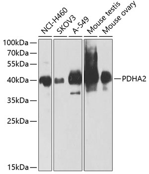
Western blot analysis of various lysates using PDHA2 Rabbit pAb (CAB9943) at 1:1000 dilution. Secondary antibody: HRP-conjugated Goat anti-Rabbit IgG (H+L) (CABS014) at 1:10000 dilution. Lysates/proteins: 25μg per lane. Blocking buffer: 3% nonfat dry milk in TBST. Detection: ECL Enhanced Kit (AbGn00021). Exposure time: 60s.
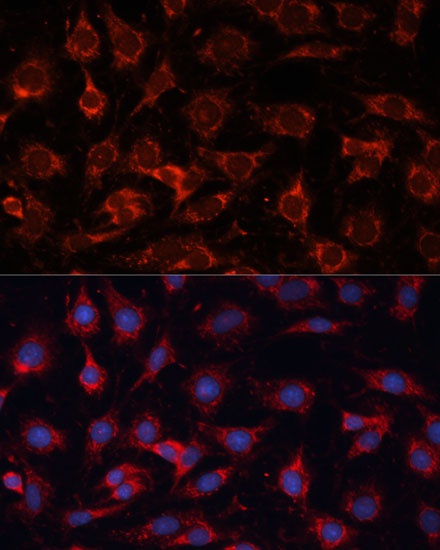
Immunofluorescence analysis of C6 cells using PDHA2 Rabbit pAb (CAB9943) at dilution of 1:100 (40x lens). Secondary antibody: Cy3-conjugated Goat anti-Rabbit IgG (H+L) (CABS007) at 1:500 dilution. Blue: DAPI for nuclear staining.
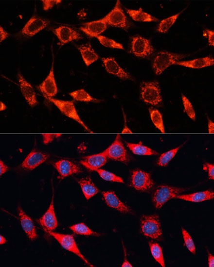
Immunofluorescence analysis of NIH-3T3 cells using PDHA2 Rabbit pAb (CAB9943) at dilution of 1:100 (40x lens). Secondary antibody: Cy3-conjugated Goat anti-Rabbit IgG (H+L) (CABS007) at 1:500 dilution. Blue: DAPI for nuclear staining.