The PDIA2 Antibody (CAB12789) is a high-quality antibody developed for reliable detection and analysis of target proteins. This antibody, raised in rabbits, exhibits high reactivity with human samples and has been validated for use in Western blot applications. By binding specifically to the PDIA2 protein, this antibody allows for accurate detection and analysis in various cell types, making it an essential component for studies in molecular biology and protein research.PDIA2, also known as protein disulfide isomerase A2, plays a crucial role in maintaining protein integrity and function by facilitating the formation and rearrangement of disulfide bonds.
This antibody is validated for use in WB, IF/ICC, ELISA applications and has demonstrated reactivity against Human, Mouse, Rat samples.
Product Name:
PDIA2 Antibody
SKU:
CAB12789
Size:
20μL, 100μL
Reactivity:
Human, Mouse, Rat
Conjugate:
Unconjugated
Immunogen:
Recombinant protein (or fragment).This information is considered to be commercially sensitive.
Recommended starting concentration is 1 μg/mL. Please optimize the concentration based on your specific assay requirements.
Synonyms:
PDI, PDA2, PDIP, PDIR, PDIA2
Positive Sample:
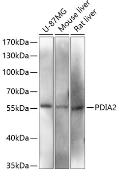
U-87MG, Mouse liver, Rat liver
Cellular Localization:
Endoplasmic Reticulum Lumen.
Calculated MW:
58kDa
Observed MW:
58kDa
This gene encodes a member of the disulfide isomerase (PDI) family of endoplasmic reticulum (ER) proteins that catalyze protein folding and thiol-disulfide interchange reactions. The encoded protein has an N-terminal ER-signal sequence, two catalytically active thioredoxin (TRX) domains, two TRX-like domains and a C-terminal ER-retention sequence. The protein plays a role in the folding of nascent proteins in the endoplasmic reticulum by forming disulfide bonds through its thiol isomerase, oxidase, and reductase activity. The encoded protein also possesses estradiol-binding activity and can modulate intracellular estradiol levels.
Purification Method
Affinity purification
Gene ID
64714
RRID
AB_2759629
Buffer Information
Store at -20℃. Avoid freeze / thaw cycles. Buffer: PBS with 0.01% thimerosal,50% glycerol,pH7.3.
Western blot analysis of various lysates using PDIA2 Rabbit pAb (CAB12789) at 1:3000 dilution. Secondary antibody: HRP-conjugated Goat anti-Rabbit IgG (H+L) (CABS014) at 1:10000 dilution. Lysates/proteins: 25μg per lane. Blocking buffer: 3% nonfat dry milk in TBST. Detection: ECL Basic Kit (AbGn00020). Exposure time: 90s.
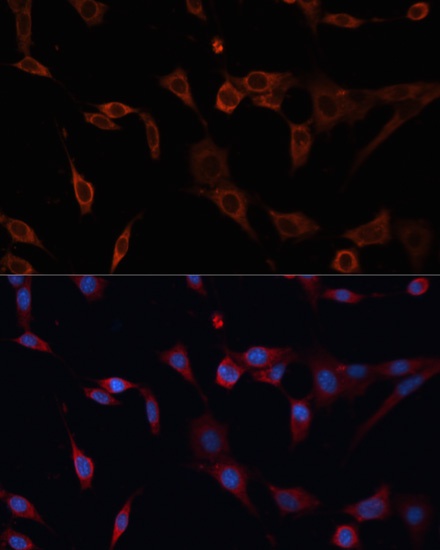
Immunofluorescence analysis of NIH/3T3 cells using PDIA2 Rabbit pAb (CAB12789) at dilution of 1:100. Secondary antibody: Cy3-conjugated Goat anti-Rabbit IgG (H+L) (CABS007) at 1:500 dilution. Blue: DAPI for nuclear staining.