The PDXDC1 Antibody (CAB14856) is a high-quality antibody developed for reliable detection and analysis of target proteins. This antibody, generated in rabbits, exhibits high specificity and sensitivity for detecting PDXDC1 in human samples, making it ideal for Western blot applications.PDXDC1, also known as pyridoxal-dependent decarboxylase domain containing 1, is a key player in pyridoxal 5'-phosphate (PLP) biosynthesis and has been implicated in cancer progression and drug resistance.
This antibody is validated for use in WB, IHC-P, ELISA applications and has demonstrated reactivity against Human, Mouse, Rat samples.
Product Name:
PDXDC1 Antibody
SKU:
CAB14856
Size:
20μL, 100μL
Reactivity:
Human, Mouse, Rat
Conjugate:
Unconjugated
Immunogen:
Recombinant protein (or fragment).This information is considered to be commercially sensitive.
Recommended starting concentration is 1 μg/mL. Please optimize the concentration based on your specific assay requirements.
Synonyms:
LP8165, PDXDC1
Positive Sample:
U-251MG, A-549, HeLa, Mouse testis, Mouse brain, Rat heart
Cellular Localization:
Golgi Apparatus.
Calculated MW:
87kDa
Observed MW:
87-106kDa
Enables cadherin binding activity. Predicted to be involved in carboxylic acid metabolic process. Located in Golgi apparatus.
Purification Method
Affinity purification
Gene ID
23042
RRID
AB_2761736
Buffer Information
Store at -20℃. Avoid freeze / thaw cycles. Buffer: PBS with 0.01% thimerosal,50% glycerol,pH7.3.
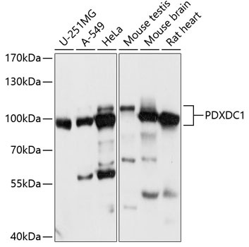
Western blot analysis of various lysates using PDXDC1 Rabbit pAb (CAB14856) at 1:1000 dilution. Secondary antibody: HRP-conjugated Goat anti-Rabbit IgG (H+L) (CABS014) at 1:10000 dilution. Lysates/proteins: 25μg per lane. Blocking buffer: 3% nonfat dry milk in TBST. Detection: ECL Basic Kit (AbGn00020). Exposure time: 30s.
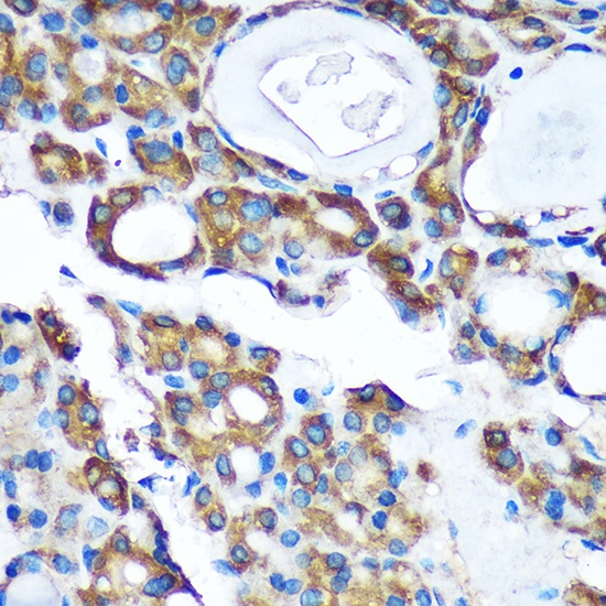
Immunohistochemistry analysis of paraffin-embedded Human thyroid cancer using PDXDC1 Rabbit pAb (CAB14856) at dilution of 1:100 (40x lens). Microwave antigen retrieval performed with 0.01M PBS Buffer (pH 7.2) prior to IHC staining.