The PEBP4 Antibody (CAB17961) is a high-quality antibody developed for reliable detection and analysis of target proteins. This rabbit polyclonal antibody is highly specific to human samples and has been validated for use in Western blot applications. By binding to the PEBP4 protein, this antibody allows for accurate detection and analysis in different cell types, making it a versatile tool for studies in cell biology and cancer research.PEBP4, also known as phosphatidylethanolamine-binding protein 4, plays a crucial role in cell signaling pathways that regulate important cellular functions.
This antibody is validated for use in WB, IHC-P, IF/ICC, ELISA applications and has demonstrated reactivity against Human, Mouse, Rat samples.
Product Name:
PEBP4 Antibody
SKU:
CAB17961
Size:
20μL, 100μL
Reactivity:
Human, Mouse, Rat
Immunogen:
Recombinant protein (or fragment).This information is considered to be commercially sensitive.
Recommended starting concentration is 1 μg/mL. Please optimize the concentration based on your specific assay requirements.
Synonyms:
PEBP-4, PEBP4
Positive Sample:
Human plasma, Rat testis
Cellular Localization:
Extracellular Region.
Calculated MW:
27kDa
Observed MW:
26kDa
Predicted to be located in extracellular region. Is expressed in bone and rib. Orthologous to human PEBP4 (phosphatidylethanolamine binding protein 4).
Purification Method
Affinity purification
Gene ID
73523
RRID
AB_2861764
Buffer Information
Store at -20℃. Avoid freeze / thaw cycles. Buffer: PBS with 0.01% thimerosal,50% glycerol,pH7.3.
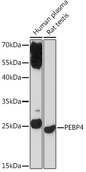
Western blot analysis of various lysates using PEBP4 Rabbit pAb (CAB17961) at 1:1000 dilution. Secondary antibody: HRP-conjugated Goat anti-Rabbit IgG (H+L) (CABS014) at 1:10000 dilution. Lysates/proteins: 25μg per lane. Blocking buffer: 3% nonfat dry milk in TBST. Detection: ECL Basic Kit (AbGn00020). Exposure time: 3min.
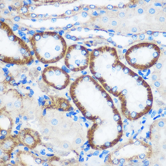
Immunohistochemistry analysis of paraffin-embedded Mouse kidney using PEBP4 Rabbit pAb (CAB17961) at dilution of 1:100 (40x lens). Microwave antigen retrieval performed with 0.01M PBS Buffer (pH 7.2) prior to IHC staining.
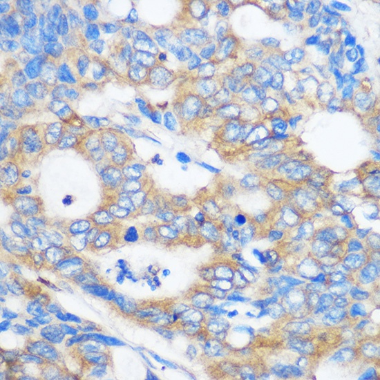
Immunohistochemistry analysis of paraffin-embedded Human colon carcinoma using PEBP4 Rabbit pAb (CAB17961) at dilution of 1:100 (40x lens). Microwave antigen retrieval performed with 0.01M PBS Buffer (pH 7.2) prior to IHC staining.
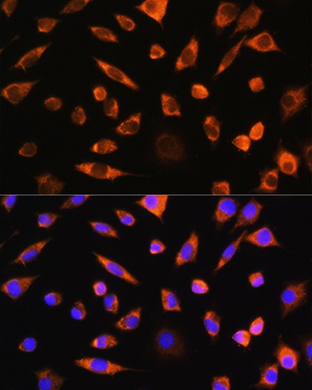
Immunofluorescence analysis of L929 cells using PEBP4 Rabbit pAb (CAB17961) at dilution of 1:100. Secondary antibody: Cy3-conjugated Goat anti-Rabbit IgG (H+L) (CABS007) at 1:500 dilution. Blue: DAPI for nuclear staining.