The PELP1 Antibody (CAB3189) is a high-quality antibody developed for reliable detection and analysis of target proteins. This antibody, produced in rabbits, demonstrates high reactivity with human samples and is validated for use in Western blot applications. By targeting the PELP1 protein, this antibody allows for the detection and analysis of PELP1 expression in different cell types, making it ideal for studies in cancer research and hormone signaling pathways.PELP1, a multifunctional protein involved in gene regulation and cell growth, is known to impact cancer cell proliferation and hormone receptor signaling.
This antibody is validated for use in WB, IHC-P, IF/ICC, ELISA applications and has demonstrated reactivity against Human, Mouse, Rat samples.
Product Name:
PELP1 Antibody
SKU:
CAB3189
Size:
20μL, 100μL
Reactivity:
Human, Mouse, Rat
Conjugate:
Unconjugated
Immunogen:
Recombinant protein (or fragment).This information is considered to be commercially sensitive.
Recommended starting concentration is 1 μg/mL. Please optimize the concentration based on your specific assay requirements.
Synonyms:
MNAR, P160, PELP1
Positive Sample:
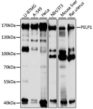
U-87MG, A-549, HeLa, NIH/3T3, Mouse liver, Rat uterus
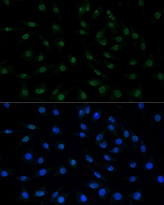
Cellular Localization:
Cytoplasm, Nucleus, Nucleoplasm.
Calculated MW:
125kDa
Observed MW:
160kDa
This gene encodes a transcription factor which coactivates transcription of estrogen receptor responsive genes and corepresses genes activated by other hormone receptors or sequence-specific transcription factors. Expression of this gene is regulated by both members of the estrogen receptor family. This gene may be involved in the progression of several types of cancer. Alternative splicing results in multiple transcript variants.
Purification Method
Affinity purification
Gene ID
27043
RRID
AB_2764975
Buffer Information
Store at -20℃. Avoid freeze / thaw cycles. Buffer: PBS with 0.01% thimerosal,50% glycerol,pH7.3.
Western blot analysis of various lysates using PELP1 Rabbit pAb (CAB3189) at 1:1000 dilution. Secondary antibody: HRP-conjugated Goat anti-Rabbit IgG (H+L) (CABS014) at 1:10000 dilution. Lysates/proteins: 25μg per lane. Blocking buffer: 3% nonfat dry milk in TBST. Detection: ECL Basic Kit (AbGn00020). Exposure time: 10s.
Immunofluorescence analysis of L929 cells using PELP1 Rabbit pAb (CAB3189) at dilution of 100 (40x lens). Secondary antibody: Cy3-conjugated Goat anti-Rabbit IgG (H+L) (CABS007) at 1:500 dilution. Blue: DAPI for nuclear staining.