The PFDN5 Antibody (CAB4014) is a high-quality antibody developed for reliable detection and analysis of target proteins. This antibody, raised in rabbits, exhibits high reactivity with human samples and has been validated for use in Western blot applications.PFDN5, also known as prefoldin subunit 5, plays a crucial role in assisting in the folding of nascent proteins, preventing their misfolding and aggregation. Its involvement in protein quality control processes makes it a target of interest for studies in molecular biology, protein chemistry, and cell biology.
This antibody is validated for use in WB, IF/ICC, ELISA applications and has demonstrated reactivity against Human, Mouse, Rat samples.
Product Name:
PFDN5 Antibody
SKU:
CAB4014
Size:
20μL, 100μL
Reactivity:
Human, Mouse, Rat
Conjugate:
Unconjugated
Immunogen:
Recombinant protein (or fragment).This information is considered to be commercially sensitive.
Recommended starting concentration is 1 μg/mL. Please optimize the concentration based on your specific assay requirements.
Synonyms:
MM1, MM-1, PFD5, PFDN5
Positive Sample:
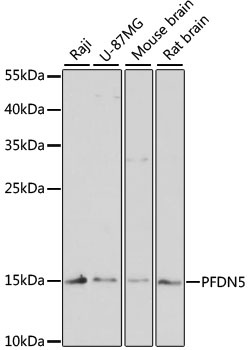
Raji, U-87MG, Mouse brain, Rat brain
Cellular Localization:
Cytoplasm, Nucleus.
Calculated MW:
17kDa
Observed MW:
17kDa
This gene encodes a member of the prefoldin alpha subunit family. The encoded protein is one of six subunits of prefoldin, a molecular chaperone complex that binds and stabilizes newly synthesized polypeptides, thereby allowing them to fold correctly. The complex, consisting of two alpha and four beta subunits, forms a double beta barrel assembly with six protruding coiled-coils. The encoded protein may also repress the transcriptional activity of the proto-oncogene c-Myc. Alternatively spliced transcript variants encoding different isoforms have been described.
Purification Method
Affinity purification
Gene ID
5204
RRID
AB_2765448
Buffer Information
Store at -20℃. Avoid freeze / thaw cycles. Buffer: PBS with 0.01% thimerosal,50% glycerol,pH7.3.
Western blot analysis of various lysates using PFDN5 Rabbit pAb (CAB4014) at 1:3000 dilution. Secondary antibody: HRP-conjugated Goat anti-Rabbit IgG (H+L) (CABS014) at 1:10000 dilution. Lysates/proteins: 25μg per lane. Blocking buffer: 3% nonfat dry milk in TBST. Detection: ECL Enhanced Kit (AbGn00021). Exposure time: 90s.
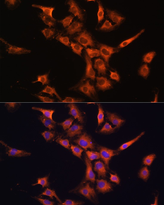
Immunofluorescence analysis of C6 cells using PFDN5 Rabbit pAb (CAB4014) at dilution of 1:100 (40x lens). Secondary antibody: Cy3-conjugated Goat anti-Rabbit IgG (H+L) (CABS007) at 1:500 dilution. Blue: DAPI for nuclear staining.
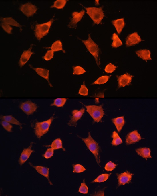
Immunofluorescence analysis of L929 cells using PFDN5 Rabbit pAb (CAB4014) at dilution of 1:100 (40x lens). Secondary antibody: Cy3-conjugated Goat anti-Rabbit IgG (H+L) (CABS007) at 1:500 dilution. Blue: DAPI for nuclear staining.