The PLGF Antibody (CAB16257) is a high-quality antibody developed for reliable detection and analysis of target proteins. This antibody, produced in rabbits, exhibits high reactivity with human samples and has been validated for use in Western blot applications. By specifically binding to the PGF protein, researchers are able to detect and analyze its expression in a variety of cell types, making it a versatile tool for studies in vascular biology, cancer research, and inflammatory diseases.PGF, also known as placental growth factor, plays a crucial role in promoting angiogenesis and inflammation, making it a potential therapeutic target for diseases such as cancer and diabetic retinopathy.
This antibody is validated for use in WB, ELISA, IF-P applications and has demonstrated reactivity against Human, Mouse, Rat samples.
Product Name:
PLGF Antibody
SKU:
CAB16257
Size:
20μL, 100μL
Reactivity:
Human, Mouse, Rat
Conjugate:
Unconjugated
Immunogen:
Recombinant protein (or fragment).This information is considered to be commercially sensitive.
Recommended starting concentration is 1 μg/mL. Please optimize the concentration based on your specific assay requirements.
Synonyms:
PGFL, PIGF, PLGF, PlGF-2, D12S1900, SHGC-10760
Positive Sample:
MCF7, Mouse placenta, Rat placenta
Cellular Localization:
Secreted.
Calculated MW:
25kDa
Observed MW:
50kDa
Enables growth factor activity. Involved in positive regulation of cell population proliferation. Predicted to be located in extracellular region. Predicted to be active in extracellular space. Implicated in several diseases, including brain ischemia; diabetic neuropathy; glioblastoma; myocardial infarction; and pancreatic endocrine carcinoma. Biomarker of several diseases, including artery disease (multiple); autoimmune disease of musculoskeletal system (multiple); epilepsy (multiple); limited scleroderma; and pancreatic endocrine carcinoma.
Purification Method
Affinity purification
Gene ID
5228
RRID
AB_2770875
Buffer Information
Store at -20℃. Avoid freeze / thaw cycles. Buffer: PBS containing 50% glycerol, preserved with proclin300 or sodium azide, pH 7.3.
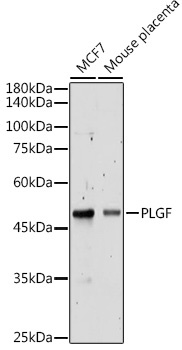
Western blot analysis of various lysates using PLGF Rabbit pAb (CAB16257) at 1:500 dilution. Secondary antibody: HRP-conjugated Goat anti-Rabbit IgG (H+L) (CABS014) at 1:10000 dilution. Lysates/proteins: 25μg per lane. Blocking buffer: 3% nonfat dry milk in TBST. Detection: ECL Basic Kit (AbGn00020). Exposure time: 30s.
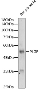
Western blot analysis of lysates from Rat placenta, using PLGF Rabbit pAb (CAB16257) at 1:500 dilution. Secondary antibody: HRP-conjugated Goat anti-Rabbit IgG (H+L) (CABS014) at 1:10000 dilution. Lysates/proteins: 25μg per lane. Blocking buffer: 3% nonfat dry milk in TBST. Detection: ECL Basic Kit (AbGn00020). Exposure time: 180s.