The PGK2 Antibody (CAB12952) is a high-quality antibody developed for reliable detection and analysis of target proteins. This antibody, produced in rabbits, exhibits high reactivity with human samples and has been validated for use in Western blot applications. By binding to the PGK2 protein, this antibody enables specific detection and analysis in a variety of cell types, making it an ideal choice for studies in biochemistry, metabolism, and cancer research.PGK2, also known as phosphoglycerate kinase 2, plays a crucial role in the energy production pathways of cells, making it essential for cellular metabolism and function. Its involvement in glycolysis and ATP production highlights its importance in processes such as cell growth, proliferation, and survival.
This antibody is validated for use in WB, IHC-P, IF/ICC, ELISA applications and has demonstrated reactivity against Human, Mouse, Rat samples.
Product Name:
PGK2 Antibody
SKU:
CAB12952
Size:
20μL, 100μL
Reactivity:
Human, Mouse, Rat
Conjugate:
Unconjugated
Immunogen:
Recombinant protein (or fragment).This information is considered to be commercially sensitive.
Recommended starting concentration is 1 μg/mL. Please optimize the concentration based on your specific assay requirements.
Synonyms:
PGKB, PGKPS, HEL-S-272, dJ417L20.2, PGK2
Positive Sample:
U-87MG, Jurkat, HeLa, HepG2, Mouse testis, Mouse brain, Mouse heart, Rat testis, Rat brain
Cellular Localization:
Cytoplasm.
Calculated MW:
45kDa
Observed MW:
45kDa
This gene is intronless, arose via retrotransposition of the phosphoglycerate kinase 1 gene, and is expressed specifically in the testis. Initially assumed to be a pseudogene, the encoded protein is actually a functional phosphoglycerate kinase that catalyzes the reversible conversion of 1,3-bisphosphoglycerate to 3-phosphoglycerate, during the Embden-Meyerhof-Parnas pathway of glycolysis, in the later stages of spermatogenesis.
Purification Method
Affinity purification
Gene ID
5232
RRID
AB_2759798
Buffer Information
Store at -20℃. Avoid freeze / thaw cycles. Buffer: PBS with 0.01% thimerosal,50% glycerol,pH7.3.
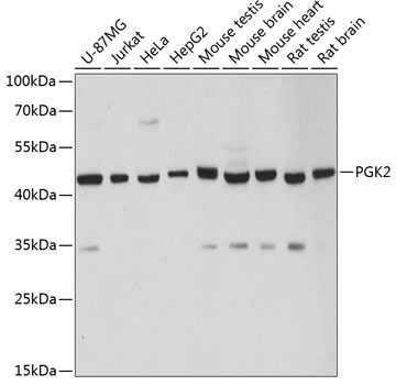
Western blot analysis of various lysates using PGK2 Rabbit pAb (CAB12952) at 1:3000 dilution. Secondary antibody: HRP-conjugated Goat anti-Rabbit IgG (H+L) (CABS014) at 1:10000 dilution. Lysates/proteins: 25μg per lane. Blocking buffer: 3% nonfat dry milk in TBST. Detection: ECL Basic Kit (AbGn00020). Exposure time: 5s.
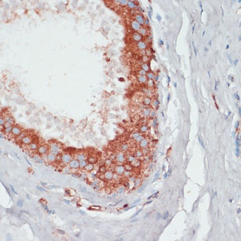
Immunohistochemistry analysis of paraffin-embedded Human mammary cancer using PGK2 Rabbit pAb (CAB12952) at dilution of 1:100 (40x lens). Microwave antigen retrieval performed with 0.01M PBS Buffer (pH 7.2) prior to IHC staining.
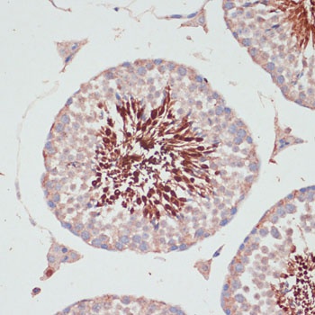
Immunohistochemistry analysis of paraffin-embedded Mouse testis using PGK2 Rabbit pAb (CAB12952) at dilution of 1:100 (40x lens). Microwave antigen retrieval performed with 0.01M PBS Buffer (pH 7.2) prior to IHC staining.
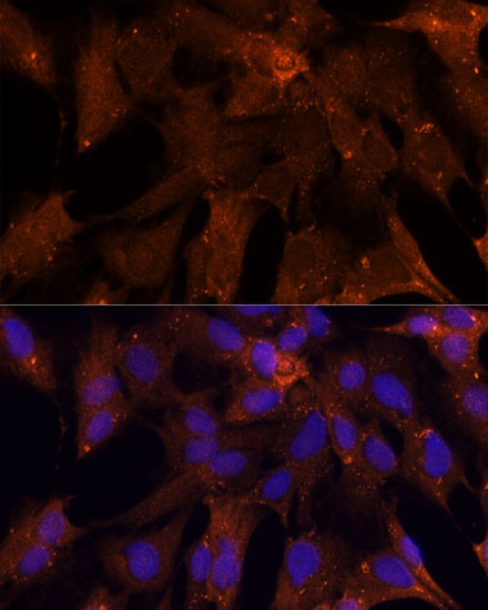
Immunofluorescence analysis of C6 cells using PGK2 Rabbit pAb (CAB12952) at dilution of 1:100 (40x lens). Secondary antibody: Cy3-conjugated Goat anti-Rabbit IgG (H+L) (CABS007) at 1:500 dilution. Blue: DAPI for nuclear staining.