The PHF11 Antibody (CAB7803) is a high-quality antibody developed for reliable detection and analysis of target proteins. This antibody, produced in rabbits, exhibits high specificity for human samples and has been validated for use in Western blotting applications. It specifically binds to the PHF11 protein, allowing for its detection and analysis in a variety of cell types.PHF11, a member of the PHD finger family of proteins, has been linked to various biological processes including transcriptional regulation, cell differentiation, and cellular proliferation.
This antibody is validated for use in WB, IHC-P, IF/ICC, ELISA applications and has demonstrated reactivity against Human, Mouse, Rat samples.
Product Name:
PHF11 Antibody
SKU:
CAB7803
Size:
20μL, 100μL
Reactivity:
Human, Mouse, Rat
Conjugate:
Unconjugated
Immunogen:
Recombinant protein (or fragment).This information is considered to be commercially sensitive.
This gene encodes a protein containing a PHD (plant homeodomain) type zinc finger. This gene has been identified in some studies as a candidate gene for asthma. Naturally-occurring readthrough transcription may occur from the upstream SETDB2 (SET domain bifurcated 2) gene to this locus. Alternative splicing results in multiple transcript variants.
Purification Method
Affinity purification
Gene ID
51131
RRID
AB_2770880
Buffer Information
Store at -20℃. Avoid freeze / thaw cycles. Buffer: PBS containing 50% glycerol, preserved with proclin300 or sodium azide, pH 7.3.
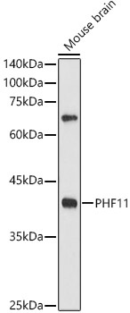
Western blot analysis of lysates from Mouse brain, using PHF11 Rabbit pAb (CAB7803) at 1:500 dilution. Secondary antibody: HRP-conjugated Goat anti-Rabbit IgG (H+L) (CABS014) at 1:10000 dilution. Lysates/proteins: 25μg per lane. Blocking buffer: 3% nonfat dry milk in TBST. Detection: ECL Enhanced Kit (AbGn00021). Exposure time: 180s.
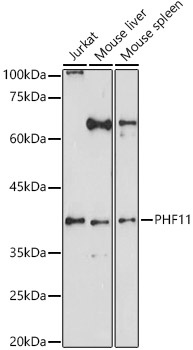
Western blot analysis of various lysates using PHF11 Rabbit pAb (CAB7803) at 1:1000 dilution. Secondary antibody: HRP-conjugated Goat anti-Rabbit IgG (H+L) (CABS014) at 1:10000 dilution. Lysates/proteins: 25μg per lane. Blocking buffer: 3% nonfat dry milk in TBST. Detection: ECL Enhanced Kit (AbGn00021). Exposure time: 30s.
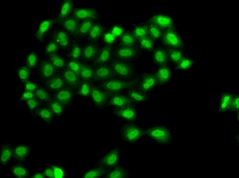
Immunofluorescence analysis of A549 cells using PHF11 Rabbit pAb (CAB7803).Secondary antibody: Cy3-conjugated Goat anti-Rabbit IgG (H+L) (CABS007) at 1:500 dilution.