The Phospho-AURKA-T288/AURKB-T232/AURKC-T198 Antibody (CABP0948) is a high-quality antibody developed for reliable detection and analysis of target proteins. This antibody, generated in rabbits, is highly specific for human samples and has been validated for use in Western blot applications. By binding to the phosphorylated forms of Aurka, Aurkb, and Aurkc, this antibody allows for the detection and analysis of these proteins in a variety of cell types.The Aurora kinases play a crucial role in the regulation of cell cycle progression, ensuring accurate chromosome segregation and cell division. Phosphorylation of these kinases at specific sites, such as T288 in Aurka, T232 in Aurkb, and T198 in Aurkc, is known to impact their activity and function.
This antibody is validated for use in WB, ELISA applications and has demonstrated reactivity against Human, Rat samples.
Product Name:
Phospho-AURKA-T288/AURKB-T232/AURKC-T198 Antibody
SKU:
CABP0948
Size:
20μL, 100μL
Reactivity:
Human, Rat
Immunogen:
Synthetic peptide. This information is considered to be commercially sensitive.
Sequence:
RTTL C
Tested Applications:
WBELISA
Recommended Dilution:
WB
1:500 - 1:2000
ELISA
Recommended starting concentration is 1 μg/mL. Please optimize the concentration based on your specific assay requirements.
The protein encoded by this gene is a cell cycle-regulated kinase that appears to be involved in microtubule formation and/or stabilization at the spindle pole during chromosome segregation. The encoded protein is found at the centrosome in interphase cells and at the spindle poles in mitosis. This gene may play a role in tumor development and progression. A processed pseudogene of this gene has been found on chromosome 1, and an unprocessed pseudogene has been found on chromosome 10. Multiple transcript variants encoding the same protein have been found for this gene. [provided by RefSeq, Jul 2008]
Purification Method
Affinity purification
Gene ID
6790 9212 6795
RRID
AB_2863858
Buffer Information
Store at -20℃. Avoid freeze / thaw cycles. Buffer: PBS with 0.01% thimerosal,50% glycerol,pH7.3.
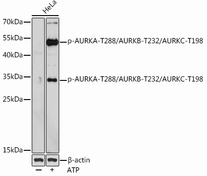
Western blot analysis of lysates from HeLa cells, using Phospho-AURKA-T288/AURKB-T232/AURKC-T198 Rabbit pAb (CABP0948) at 1:1000 dilution. Hela cells were treated with ATP(5 mM) at 30℃ for 1 hour. Secondary antibody: HRP-conjugated Goat anti-Rabbit IgG (H+L) (CABS014) at 1:10000 dilution. Lysates/proteins: 25μg per lane. Blocking buffer: 3% BSA. Detection: ECL Basic Kit (AbGn00020). Exposure time: 90s.