The Phospho-Bcl-2-S70 Antibody (CABP0575) is a high-quality antibody developed for reliable detection and analysis of target proteins. The antibody, raised in rabbits, is highly specific for detecting phosphorylated BCL2 in human samples, making it ideal for Western blot applications.Phosphorylation of BCL2 at serine 70 has been shown to play a role in regulating cell survival and apoptosis, making it a critical target for cancer research and drug development. By targeting this specific phosphorylation site, researchers can gain insights into the mechanisms underlying BCL2-mediated cell death and survival pathways.
This antibody is validated for use in WB, IF/ICC, ELISA applications and has demonstrated reactivity against Human, Rat samples.
Product Name:
Phospho-Bcl-2-S70 Antibody
SKU:
CABP0575
Size:
20μL, 100μL
Reactivity:
Human, Rat
Conjugate:
Unconjugated
Immunogen:
Synthetic peptide. This information is considered to be commercially sensitive.
Sequence:
RTSP L
Tested Applications:
WBIF/ICCELISA
Recommended Dilution:
WB
1:500 - 1:2000
IF/ICC
1:50 - 1:200
ELISA
Recommended starting concentration is 1 μg/mL. Please optimize the concentration based on your specific assay requirements.
This gene encodes an integral outer mitochondrial membrane protein that blocks the apoptotic death of some cells such as lymphocytes. Constitutive expression of BCL2, such as in the case of translocation of BCL2 to Ig heavy chain locus, is thought to be the cause of follicular lymphoma. Alternative splicing results in multiple transcript variants.
Purification Method
Affinity purification
Gene ID
596
RRID
AB_2736880
Buffer Information
Store at -20℃. Avoid freeze / thaw cycles. Buffer: PBS containing 50% glycerol, preserved with proclin300 or sodium azide, pH 7.3.
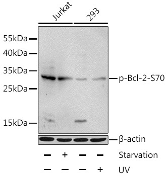
Western blot analysis of lysates from Jurkat and 293 cells, using Phospho-Bcl-2-S70 Rabbit pAb (CABP0575) at 1:1000 dilution. Jurkat cells were treated by serum-starvation overnight. 293 cells were treated by UV for 15-30 minutes. Secondary antibody: HRP-conjugated Goat anti-Rabbit IgG (H+L) (CABS014) at 1:10000 dilution. Lysates/proteins: 25μg per lane. Blocking buffer: 3% BSA.
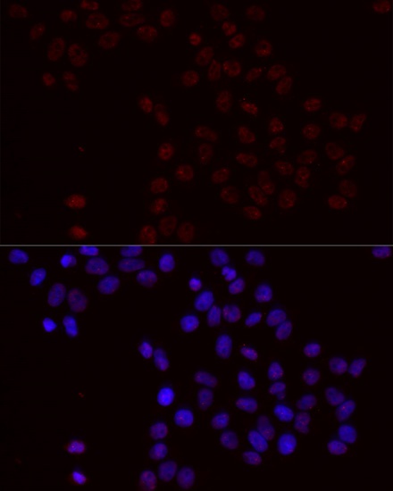
Immunofluorescence analysis of HeLa cells using Phospho-Bcl-2-S70 Rabbit pAb (CABP0575) at dilution of 1:100 (40x lens). Secondary antibody: Cy3-conjugated Goat anti-Rabbit IgG (H+L) (CABS007) at 1:500 dilution. Blue: DAPI for nuclear staining.