The BCR Antibody (CAB0068) is a high-quality antibody developed for reliable detection and analysis of target proteins. This antibody, generated in rabbits, exhibits high reactivity with human samples and is validated for use in Western blot applications. By targeting the BCR protein, this antibody enables accurate detection and analysis in a variety of cell types, making it an essential component for studies in immunology and cancer research.
This antibody is validated for use in WB, IHC-P, ELISA applications and has demonstrated reactivity against Human, Mouse, Rat samples.
Product Name:
BCR Antibody
SKU:
CAB0068
Size:
20μL, 100μL
Reactivity:
Human, Mouse, Rat
Conjugate:
Unconjugated
Immunogen:
Recombinant protein (or fragment).This information is considered to be commercially sensitive.
A reciprocal translocation between chromosomes 22 and 9 produces the Philadelphia chromosome, which is often found in patients with chronic myelogenous leukemia. The chromosome 22 breakpoint for this translocation is located within the BCR gene. The translocation produces a fusion protein which is encoded by sequence from both BCR and ABL, the gene at the chromosome 9 breakpoint. Although the BCR-ABL fusion protein has been extensively studied, the function of the normal BCR gene product is not clear. The unregulated tyrosine kinase activity of BCR-ABL1 contributes to the immortality of leukaemic cells. The BCR protein has serine/threonine kinase activity and is a GTPase-activating protein for p21rac and other kinases. Two transcript variants encoding different isoforms have been found for this gene.
Purification Method
Affinity purification
Gene ID
613
RRID
AB_2756929
Buffer Information
Store at -20℃. Avoid freeze / thaw cycles. Buffer: PBS containing 50% glycerol, preserved with proclin300 or sodium azide, pH 7.3.
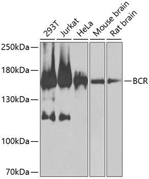
Western blot analysis of various lysates using BCR Rabbit pAb (CAB0068) at 1:1000 dilution. Secondary antibody: HRP-conjugated Goat anti-Rabbit IgG (H+L) (CABS014) at 1:10000 dilution. Lysates/proteins: 25μg per lane. Blocking buffer: 3% nonfat dry milk in TBST. Detection: ECL Basic Kit (AbGn00020). Exposure time: 30s.
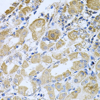
Immunohistochemistry analysis of paraffin-embedded Human stomach using BCR Rabbit pAb (CAB0068) at dilution of 1:100 (40x lens). Microwave antigen retrieval performed with 0.01M PBS Buffer (pH 7.2) prior to IHC staining.