The Phospho-c-Jun-T91 Monoclonal Antibody (CABP1003) is a high-quality antibody developed for reliable detection and analysis of target proteins. This antibody, raised in rabbits, is highly reactive with human samples and has been validated for use in Western blot applications.The phosphorylation of c-Jun at threonine 91 plays a crucial role in various cellular processes, including cell growth, differentiation, and apoptosis. By targeting this specific phosphorylation site, researchers can gain insights into the signaling pathways and mechanisms involved in these processes.
This antibody is validated for use in WB, IHC-P, ELISA applications and has demonstrated reactivity against Human, Mouse samples.
Product Name:
Phospho-c-Jun-T91 Monoclonal Antibody
SKU:
CABP1003
Size:
20μL, 100μL
Reactivity:
Human, Mouse
Clone Number:
ARC1548
Conjugate:
Unconjugated
Immunogen:
Synthetic peptide. This information is considered to be commercially sensitive.
Sequence:
TTTP T
Tested Applications:
WBIHC-PELISA
Recommended Dilution:
WB
1:500 - 1:2000
IHC-P
1:50 - 1:200
ELISA
Recommended starting concentration is 1 μg/mL. Please optimize the concentration based on your specific assay requirements.
Synonyms:
AP1, p39, AP-1, cJUN, c-Jun, Phospho-c-Jun-T91
Positive Sample:
NIH/3T3 treated with UV, NIH/3T3 treated with Anisomycin, 293T treated with UV
Cellular Localization:
Nucleus.
Calculated MW:
36kDa
Observed MW:
48kDa
This gene is the putative transforming gene of avian sarcoma virus 17. It encodes a protein which is highly similar to the viral protein, and which interacts directly with specific target DNA sequences to regulate gene expression. This gene is intronless and is mapped to 1p32-p31, a chromosomal region involved in both translocations and deletions in human malignancies.
Purification Method
Affinity purification
Gene ID
3725
RRID
AB_2863895
Buffer Information
Store at -20℃. Avoid freeze / thaw cycles. Buffer: PBS containing 50% glycerol and 0.05% BSA, preserved with proclin300 or sodium azide, pH 7.3.
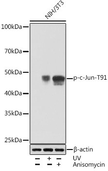
Western blot analysis of lysates from NIH/3T3 cells, using Phospho-c-Jun-T91 Rabbit mAb (CABP1003) at 1:1000 dilution. NIH/3T3 cells were treated with UV at room temperature for 15-30 minutes or treated with Anisomycin (25 μg/mL) at 37℃ for 30 minutes. Secondary antibody: HRP-conjugated Goat anti-Rabbit IgG (H+L) (CABS014) at 1:10000 dilution. Lysates/proteins: 25μg per lane. Blocking buffer: 3% BSA. Detection: ECL Basic Kit (AbGn00020). Exposure time: 30s.
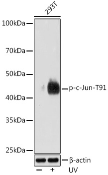
Western blot analysis of lysates from 293T cells, using Phospho-c-Jun-T91 Rabbit mAb (CABP1003) at 1:1000 dilution. 293T cells were treated with UV at room temperature for 15-30 minutes. Secondary antibody: HRP-conjugated Goat anti-Rabbit IgG (H+L) (CABS014) at 1:10000 dilution. Lysates/proteins: 25μg per lane. Blocking buffer: 3% BSA. Detection: ECL Enhanced Kit (AbGn00021). Exposure time: 3min.