The Phospho-CDK2-T160 Antibody (CABP0325) is a high-quality antibody developed for reliable detection and analysis of target proteins. This antibody is raised in rabbits and specifically targets the phosphorylated form of CDK2 at threonine 160, allowing for detection and analysis in various cell types.CDK2 is a key regulator of cell cycle progression, with phosphorylation at threonine 160 playing an important role in its activity. By targeting this specific phosphorylation site, researchers can gain insights into the mechanisms underlying cell cycle control and potentially identify novel therapeutic targets for diseases like cancer.
This antibody is validated for use in WB, IHC-P, ELISA applications and has demonstrated reactivity against Human, Mouse, Rat samples.
Product Name:
Phospho-CDK2-T160 Antibody
SKU:
CABP0325
Size:
20μL, 100μL
Reactivity:
Human, Mouse, Rat
Conjugate:
Unconjugated
Immunogen:
Synthetic peptide. This information is considered to be commercially sensitive.
Sequence:
TYTH E
Tested Applications:
WBIHC-PELISA
Recommended Dilution:
WB
1:500 - 1:5000
IHC-P
1:50 - 1:100
ELISA
Recommended starting concentration is 1 μg/mL. Please optimize the concentration based on your specific assay requirements.
This gene encodes a member of a family of serine/threonine protein kinases that participate in cell cycle regulation. The encoded protein is the catalytic subunit of the cyclin-dependent protein kinase complex, which regulates progression through the cell cycle. Activity of this protein is especially critical during the G1 to S phase transition. This protein associates with and regulated by other subunits of the complex including cyclin A or E, CDK inhibitor p21Cip1 (CDKN1A), and p27Kip1 (CDKN1B). Alternative splicing results in multiple transcript variants.
Purification Method
Affinity purification
Gene ID
1017
RRID
AB_2770980
Buffer Information
Store at -20℃. Avoid freeze / thaw cycles. Buffer: PBS containing 50% glycerol, preserved with proclin300 or sodium azide, pH 7.3.
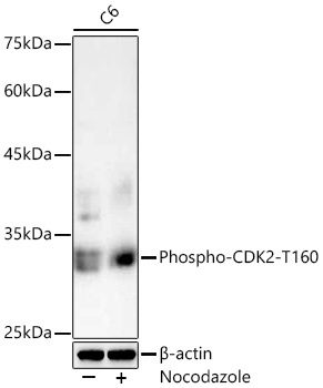
Western blot analysis of lysates from C6 cells using Phospho-CDK2-T160 Rabbit pAb (CABP0325) at 1:1000 dilution. C6 cells were treated with Nocodazole (10 ng/ml) at 37℃ for 20 hours. Secondary antibody: HRP-conjugated Goat anti-Rabbit IgG (H+L) (CABS014) at 1:10000 dilution. Lysates/proteins: 20 μg per lane. Blocking buffer: 3% nonfat dry milk in TBST. Detection: ECL Basic Kit (AbGn00020). Exposure time: 3s.
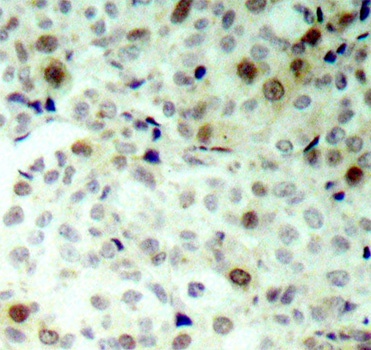
Immunohistochemistry analysis of paraffin-embedded Human breast carcinoma using Phospho-CDK2-T160 Rabbit pAb (CABP0325). Microwave antigen retrieval performed with 0.01M Tris/EDTA Buffer (pH 9.0) prior to IHC staining.