The Phospho-Chk1-S345 Antibody (CABP0578) is a high-quality antibody developed for reliable detection and analysis of target proteins. Chek1 is a key regulator of cell cycle checkpoints and DNA damage response pathways. This antibody, raised in rabbits, exhibits high reactivity with human samples and has been validated for use in Western blot applications.Targeting the phosphorylation site at serine 345 of Chek1, this antibody allows for the detection and analysis of Chek1 activation in various cell types, providing valuable insights into cell cycle regulation, DNA damage repair, and cancer biology.
This antibody is validated for use in WB, ELISA applications and has demonstrated reactivity against Human, Mouse, Rat samples.
Product Name:
Phospho-Chk1-S345 Antibody
SKU:
CABP0578
Size:
20μL, 100μL
Reactivity:
Human, Mouse, Rat
Conjugate:
Unconjugated
Immunogen:
Synthetic peptide. This information is considered to be commercially sensitive.
Sequence:
SFSQ P
Tested Applications:
WBELISA
Recommended Dilution:
WB
1:100 - 1:500
ELISA
Recommended starting concentration is 1 μg/mL. Please optimize the concentration based on your specific assay requirements.
The protein encoded by this gene belongs to the Ser/Thr protein kinase family. It is required for checkpoint mediated cell cycle arrest in response to DNA damage or the presence of unreplicated DNA. This protein acts to integrate signals from ATM and ATR, two cell cycle proteins involved in DNA damage responses, that also associate with chromatin in meiotic prophase I. Phosphorylation of CDC25A protein phosphatase by this protein is required for cells to delay cell cycle progression in response to double-strand DNA breaks. Several alternatively spliced transcript variants have been found for this gene.
Purification Method
Affinity purification
Gene ID
1111
RRID
AB_2770995
Buffer Information
Store at -20℃. Avoid freeze / thaw cycles. Buffer: PBS containing 50% glycerol, preserved with proclin300 or sodium azide, pH 7.3.
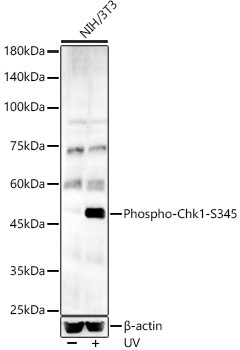
Western blot analysis of lysates from NIH/3T3 cells, using Phospho-Chk1-S345 Rabbit pAb (CABP0578) at 1:500 dilution. NIH/3T3 cells were treated with UV at room temperature for 15-30 minutes. Secondary antibody: HRP-conjugated Goat anti-Rabbit IgG (H+L) (CABS014) at 1:10000 dilution. Lysates/proteins: 25μg per lane. Blocking buffer: 3% nonfat dry milk in TBST. Detection: ECL Basic Kit (AbGn00020). Exposure time: 180s.