The DAB1 Antibody (CAB10349) is a high-quality antibody developed for reliable detection and analysis of target proteins. This antibody, generated in rabbits, exhibits high specificity and sensitivity towards Disabled Homolog 1 in human samples, making it suitable for use in Western blotting and immunohistochemistry applications.Disabled Homolog 1, also referred to as DAB1, is a key player in neuronal development and synaptic plasticity, making it a compelling target for neuroscience and neurodevelopmental research.
This antibody is validated for use in WB, IF/ICC, ELISA applications and has demonstrated reactivity against Human, Mouse, Rat samples.
Product Name:
DAB1 Antibody
SKU:
CAB10349
Size:
20μL, 100μL
Reactivity:
Human, Mouse, Rat
Conjugate:
Unconjugated
Immunogen:
Recombinant protein (or fragment).This information is considered to be commercially sensitive.
Recommended starting concentration is 1 μg/mL. Please optimize the concentration based on your specific assay requirements.
Synonyms:
SCA37, DAB1
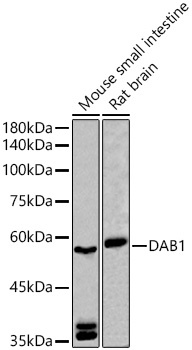
Positive Sample:
Mouse small intestine, Rat brain
Cellular Localization:
Cytoplasm, Cytosol, Perinuclear Region Of Cytoplasm.
Calculated MW:
64kDa
Observed MW:
57kDa/59kDa
The laminar organization of multiple neuronal types in the cerebral cortex is required for normal cognitive function. In mice, the disabled-1 gene plays a central role in brain development, directing the migration of cortical neurons past previously formed neurons to reach their proper layer. This gene is similar to disabled-1, and the protein encoded by this gene is thought to be a signal transducer that interacts with protein kinase pathways to regulate neuronal positioning in the developing brain.
Purification Method
Affinity purification
Gene ID
1600
RRID
AB_2757894
Buffer Information
Store at -20℃. Avoid freeze / thaw cycles. Buffer: PBS containing 50% glycerol, preserved with proclin300 or sodium azide, pH 7.3.
Western blot analysis of various lysates using DAB1 Rabbit pAb (CAB10349) at 1:500 dilution. Secondary antibody: HRP-conjugated Goat anti-Rabbit IgG (H+L) (CABS014) at 1:10000 dilution. Lysates/proteins: 25μg per lane. Blocking buffer: 3% nonfat dry milk in TBST. Detection: ECL Basic Kit (AbGn00020). Exposure time: 90s.
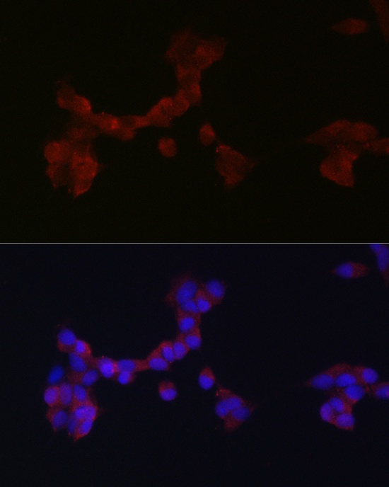
Immunofluorescence analysis of SH-SY5Y cells using DAB1 Rabbit pAb (CAB10349) at dilution of 1:200 (40x lens). Secondary antibody: Cy3-conjugated Goat anti-Rabbit IgG (H+L) (CABS007) at 1:500 dilution. Blue: DAPI for nuclear staining.