The Phospho-ERK1-T202 + ERK2-T185 Monoclonal Antibody (CABP0485) is a high-quality antibody developed for reliable detection and analysis of target proteins. This antibody, generated in rabbits, is highly specific for detecting phosphorylated ERK1 at threonine 202 and phosphorylated ERK2 at threonine 185 in human samples.The antibody is validated for use in Western blot applications, allowing for precise detection and quantification of phosphorylated ERK1 and ERK2 levels in various cell types.
This antibody is validated for use in WB, IHC-P, ELISA applications and has demonstrated reactivity against Human, Mouse, Rat samples.
Product Name:
Phospho-ERK1-T202 + ERK2-T185 Monoclonal Antibody
SKU:
CABP0485
Size:
20μL, 100μL
Reactivity:
Human, Mouse, Rat
Clone Number:
ARC0100
Conjugate:
Unconjugated
Immunogen:
Synthetic peptide. This information is considered to be commercially sensitive.
Sequence:
FLTE Y
Tested Applications:
WBIHC-PELISA
Recommended Dilution:
WB
1:500 - 1:2000
IHC-P
1:50 - 1:200
ELISA
Recommended starting concentration is 1 μg/mL. Please optimize the concentration based on your specific assay requirements.
Caveola, Cytoplasm, Cytoskeleton, Cytosol, Early Endosome, Endoplasmic Reticulum Lumen, Extracellular Region, Focal Adhesion, Golgi Apparatus, Late Endosome, Microtubule Organizing Center, Mitochondrion, Mitotic Spindle, Nucleoplasm, Nucleus, Plasma Membrane.
Calculated MW:
42,44kDa
Observed MW:
42kDa/44kDa
Histones are basic nuclear proteins that are responsible for the nucleosome structure of the chromosomal fiber in eukaryotes. Nucleosomes consist of approximately 146 bp of DNA wrapped around a histone octamer composed of pairs of each of the four core histones (H2A, H2B, H3, and H4). The chromatin fiber is further compacted through the interaction of a linker histone, H1, with the DNA between the nucleosomes to form higher order chromatin structures. This gene is intronless and encodes a replication-dependent histone that is a member of the histone H3 family. Transcripts from this gene lack polyA tails; instead, they contain a palindromic termination element. This gene is located separately from the other H3 genes that are in the histone gene cluster on chromosome 6p22-p21.3.
Purification Method
Affinity purification
Gene ID
5594 5595
RRID
AB_2863806
Buffer Information
Store at -20℃. Avoid freeze / thaw cycles. Buffer: PBS containing 50% glycerol and 0.05% BSA, preserved with proclin300 or sodium azide, pH 7.3.
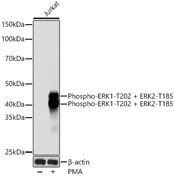
Western blot analysis of lysates from Jurkat cells, using Phospho-ERK1-T202 + ERK2-T185 Rabbit mAb (CABP0485) at 1:1000 dilution. Jurkat cells were treated with PMA/TPA (200 nM) at 37℃ for 10 minutes. Secondary antibody: HRP-conjugated Goat anti-Rabbit IgG (H+L) (CABS014) at 1:10000 dilution. Lysates/proteins: 25μg per lane. Blocking buffer: 3% nonfat dry milk in TBST. Detection: ECL Basic Kit (AbGn00020). Exposure time: 10s.
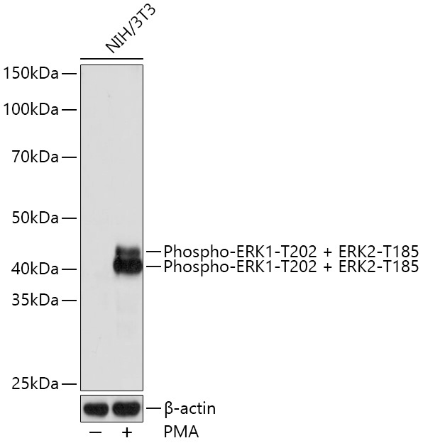
Western blot analysis of lysates from NIH/3T3 cells, using Phospho-ERK1-T202 + ERK2-T185 Rabbit mAb (CABP0485) at 1:1000 dilution. NIH/3T3 cells were treated with PMA/TPA (200 nM) at 37℃ for 30 minutes after serum-starvation overnight. Secondary antibody: HRP-conjugated Goat anti-Rabbit IgG (H+L) (CABS014) at 1:10000 dilution. Lysates/proteins: 25μg per lane. Blocking buffer: 3% nonfat dry milk in TBST. Detection: ECL Basic Kit (AbGn00020). Exposure time: 1S.
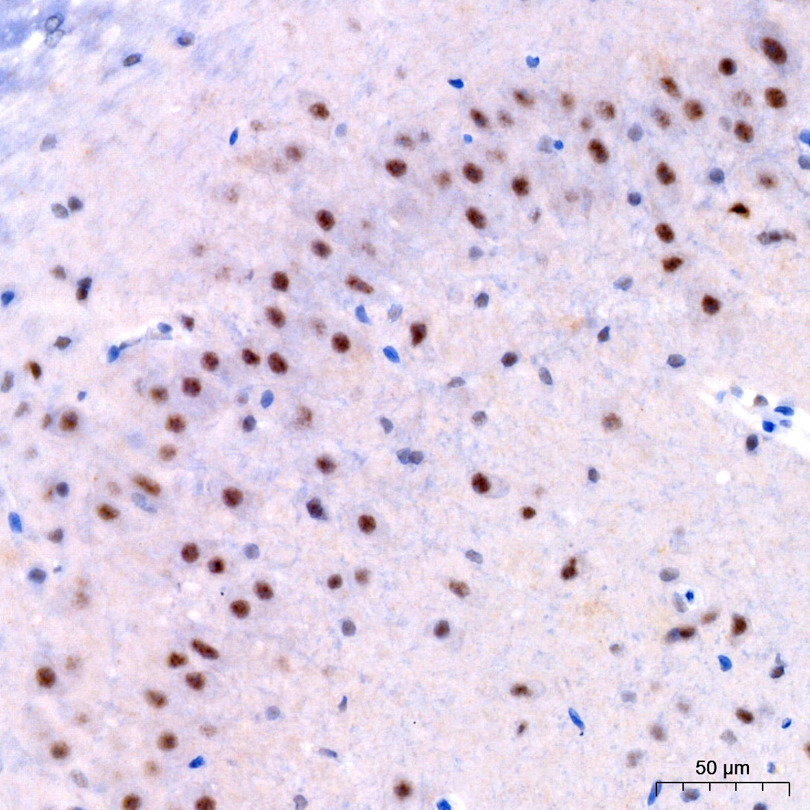
Immunohistochemistry analysis of paraffin-embedded Rat brain using Phospho-ERK1-T202 + ERK2-T185 Rabbit mAb (CABP0485) at dilution of 1:200 (40x lens). High pressure antigen retrieval performed with 0.01M Citrate buffer (pH 6.0) prior to IHC staining.