The Phospho-GSK3beta-S9 Antibody (CABP0039) is a high-quality antibody developed for reliable detection and analysis of target proteins. This antibody, produced in rabbits, is highly specific for human samples and has been validated for use in Western blot applications. By targeting the phosphorylated form of GSK3B, this antibody allows for the detection and analysis of GSK3B activation in different cell types, making it an essential reagent for investigations in cell signaling, metabolism, and cancer biology.
This antibody is validated for use in WB, IHC-P, IF/ICC, ELISA applications and has demonstrated reactivity against Human, Mouse, Rat samples.
Product Name:
Phospho-GSK3beta-S9 Antibody
SKU:
CABP0039
Size:
20μL, 100μL
Reactivity:
Human, Mouse, Rat
Conjugate:
Unconjugated
Immunogen:
Synthetic peptide. This information is considered to be commercially sensitive.
Sequence:
TTSF AE
Tested Applications:
WBIHC-PIF/ICCELISA
Recommended Dilution:
WB
1:500 - 1:5000
IF/ICC
1:50 - 1:200
IHC-P
1:50 - 1:200
ELISA
Recommended starting concentration is 1 μg/mL. Please optimize the concentration based on your specific assay requirements.
Synonyms:
GSK3B, gsk-3β, Phospho-GSK3β-S9
Positive Sample:
C6 treated with Calyculin A
Cellular Localization:
Cell Membrane, Cytoplasm, Nucleus.
Calculated MW:
47kDa
Observed MW:
46kDa
The protein encoded by this gene is a serine-threonine kinase belonging to the glycogen synthase kinase subfamily. It is a negative regulator of glucose homeostasis and is involved in energy metabolism, inflammation, ER-stress, mitochondrial dysfunction, and apoptotic pathways. Defects in this gene have been associated with Parkinson disease and Alzheimer disease.
Purification Method
Affinity purification
Gene ID
2932
RRID
AB_2771151
Buffer Information
Store at -20℃. Avoid freeze / thaw cycles. Buffer: PBS containing 50% glycerol, preserved with proclin300 or sodium azide, pH 7.3.
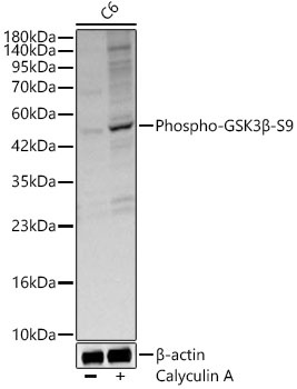
Western blot analysis of lysates from C6 cells using Phospho-GSK3β-S9 Rabbit pAb (CABP0039) at 1:500 dilution. C6 cells were treated with Calyculin A (100 nM) at 37℃ for 30 minutes after serum-starvation overnight. Secondary antibody: HRP-conjugated Goat anti-Rabbit IgG (H+L) (CABS014) at 1:10000 dilution. Lysates/proteins: 25 μg per lane. Blocking buffer: 3% nonfat dry milk in TBST. Detection: ECL Basic Kit (AbGn00020). Exposure time: 10s.
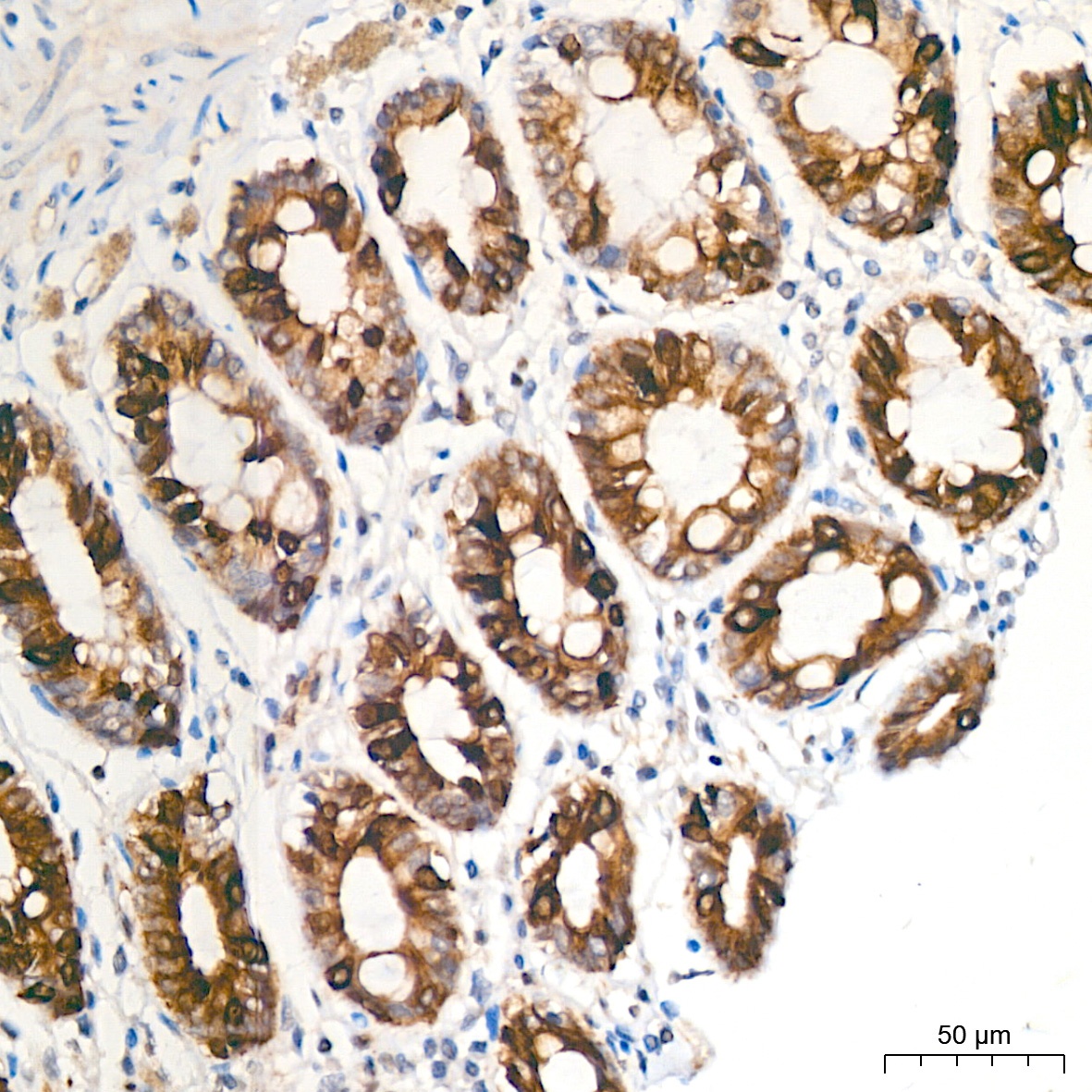
Immunohistochemistry analysis of paraffin-embedded Human colon tissue using Phospho-GSK3β-S9 Rabbit pAb (CABP0039) at a dilution of 1:100 (40x lens). High pressure antigen retrieval was performed with 0.01 M citrate buffer (pH 6.0) prior to IHC staining.
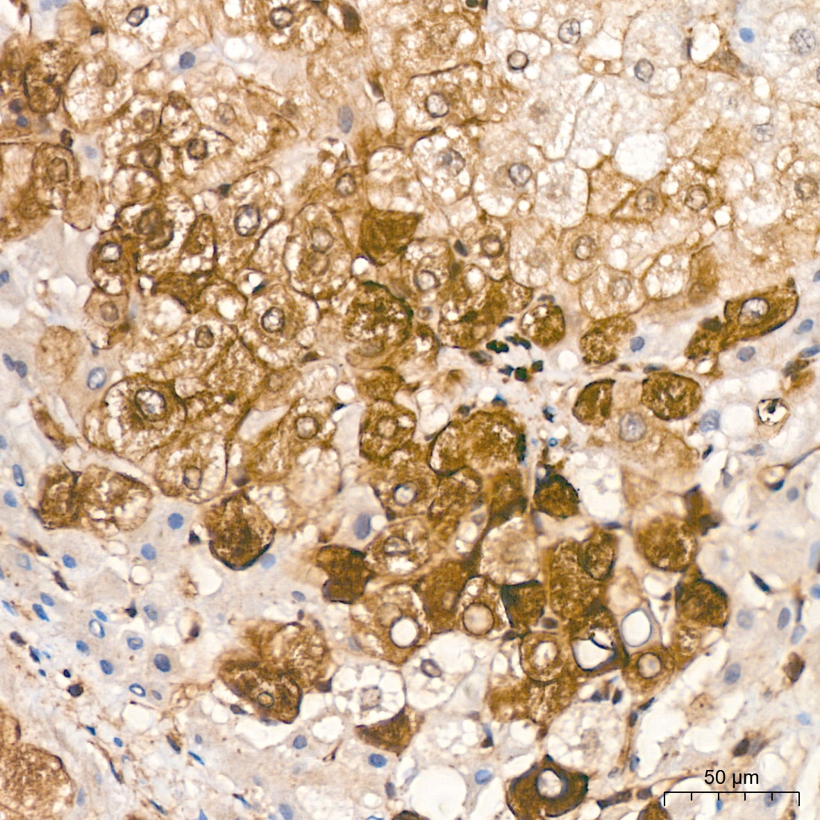
Immunohistochemistry analysis of paraffin-embedded Human liver cancer tissue using Phospho-GSK3β-S9 Rabbit pAb (CABP0039) at a dilution of 1:100 (40x lens). High pressure antigen retrieval was performed with 0.01 M citrate buffer (pH 6.0) prior to IHC staining.
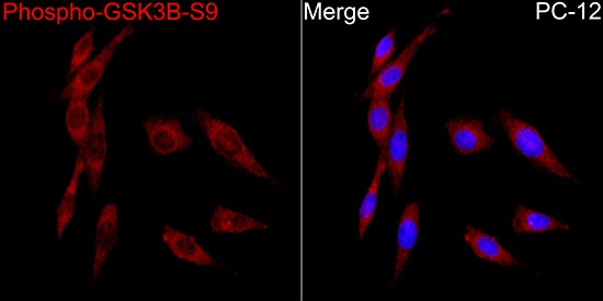
Immunofluorescence analysis of PC-12 cells using Phospho-GSK3β-S9 Rabbit pAb(CABP0039) at a dilution of 1:100 (40x lens). Secondary antibody:Cy3 Goat Anti-Rabbit IgG (H+L)(CABS007) at 1:500 dilution. Blue: DAPI for nuclear staining.
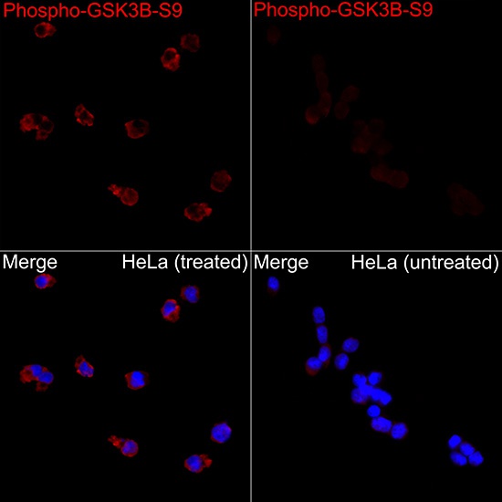
Immunofluorescence analysis of HeLa CA and HeLa cells using Phospho-GSK3β-S9 Rabbit pAb(CABP0039) at a dilution of 1:100 (40x lens). Secondary antibody:Cy3 Goat Anti-Rabbit IgG (H+L)(CABS007) at 1:500 dilution. Blue: DAPI for nuclear staining.