The Phospho-HSL-S660 Monoclonal Antibody (CABP1432) is a high-quality antibody developed for reliable detection and analysis of target proteins. HSL is a key enzyme involved in the regulation of lipid metabolism and plays a crucial role in the mobilization of stored fats in the body.This monoclonal antibody, produced through advanced technology, is highly reactive with phosphorylated HSL at serine 660 in human, mouse, and rat samples. It is validated for use in various applications including Western blot, immunohistochemistry, and immunofluorescence, allowing for precise detection and analysis of phospho-HSL (S660) in different cell types and tissues.
This antibody is validated for use in WB, ELISA applications and has demonstrated reactivity against Mouse samples.
Product Name:
Phospho-HSL-S660 Monoclonal Antibody
SKU:
CABP1432
Size:
20μL, 100μL
Reactivity:
Mouse
Clone Number:
ARC61254
Conjugate:
Unconjugated
Immunogen:
Synthetic peptide. This information is considered to be commercially sensitive.
Tested Applications:
WBELISA
Recommended Dilution:
WB
1:500 - 1:1000
ELISA
Recommended starting concentration is 1 μg/mL. Please optimize the concentration based on your specific assay requirements.
The protein encoded by this gene has a long and a short form, generated by use of alternative translational start codons. The long form is expressed in steroidogenic tissues such as testis, where it converts cholesteryl esters to free cholesterol for steroid hormone production. The short form is expressed in adipose tissue, among others, where it hydrolyzes stored triglycerides to free fatty acids.
Purification Method
Affinity purification
Gene ID
3991
Buffer Information
Store at -20℃. Avoid freeze / thaw cycles. Buffer: PBS containing 50% glycerol and 0.05% BSA, preserved with proclin300 or sodium azide, pH 7.3.
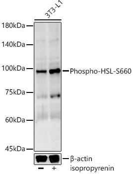
Western blot analysis of lysates from 3T3-L1 cells, using Phospho-HSL-S660 Rabbit mAb (CABP1432) at 1:1000 dilution. 3T3-L1 cells that were not differentiated or differentiated into adipocytes treated with 0. 06mol/L alcohol and treated with isoproterenol (10 μM, 20 min). Secondary antibody: HRP-conjugated Goat anti-Rabbit IgG (H+L) (CABS014) at 1:10000 dilution. Lysates/proteins: 25μg per lane. Blocking buffer: 3% nonfat dry milk in TBST. Detection: ECL Basic Kit (AbGn00020). Exposure time: 180s.
 at 1:1000 dilution. 3T3-L1 cells that were not differentiated or differentiated into adipocytes treated with 0. 06mol/L alcohol and treated with isoproterenol (10 μM, 20 min). Secondary antibody: HRP Goat Anti-Rabbit IgG (H+L) at 1:10000 dilution. Lysates/proteins: 25μg per lane. Blocking buffer: 3% nonfat dry milk in TBST.")
 at 1:1000 dilution. 3T3-L1 cells that were not differentiated or differentiated into adipocytes treated with 0. 06mol/L alcohol and treated with isoproterenol (10 μM, 20 min). Secondary antibody: HRP Goat Anti-Rabbit IgG (H+L) at 1:10000 dilution. Lysates/proteins: 25μg per lane. Blocking buffer: 3% nonfat dry milk in TBST.")


 at 37℃ for 30 minutes after serum-starvation overnight. HeLa cells were treated by nocodazole (50 ng/ml) at 37℃ for 20 hours. Secondary antibody: HRP Goat Anti-Rabbit IgG (H+L) at 1:10000 dilution. Lysates/proteins: 25ug per lane. Blocking buffer: 3% nonfat dry milk in TBST. Detection: ECL Enhanced Kit. Exposure time: 300s.")
