The Phospho-IRF3-S386 Monoclonal Antibody (CABP0995) is a high-quality antibody developed for reliable detection and analysis of target proteins. This antibody, generated in rabbits, specifically targets the phosphorylated form of IRF3 at serine 386, a key post-translational modification involved in the activation of innate immune responses.Validated for use in Western blotting and immunohistochemistry applications, the Phospho-IRF3 (S386) antibody allows for the detection and quantification of phosphorylated IRF3 in various cell types and tissues.
This antibody is validated for use in WB, ELISA applications and has demonstrated reactivity against Human samples.
Product Name:
Phospho-IRF3-S386 Monoclonal Antibody
SKU:
CABP0995
Size:
20μL, 100μL
Reactivity:
Human
Clone Number:
ARC1539
Conjugate:
Unconjugated
Immunogen:
Synthetic peptide. This information is considered to be commercially sensitive.
Sequence:
ASSL E
Tested Applications:
WBELISA
Recommended Dilution:
WB
1:500 - 1:2000
ELISA
Recommended starting concentration is 1 μg/mL. Please optimize the concentration based on your specific assay requirements.
Synonyms:
IIAE7, Phospho-IRF3-S386
Positive Sample:
HeLa treated with Calyculin A
Cellular Localization:
Cytoplasm, Nucleus.
Calculated MW:
47kDa
Observed MW:
55kDa
This gene encodes a member of the interferon regulatory transcription factor (IRF) family. The encoded protein is found in an inactive cytoplasmic form that upon serine/threonine phosphorylation forms a complex with CREBBP. This complex translocates to the nucleus and activates the transcription of interferons alpha and beta, as well as other interferon-induced genes. The protein plays an important role in the innate immune response against DNA and RNA viruses. Mutations in this gene are associated with Encephalopathy, acute, infection-induced, herpes-specific, 7.
Purification Method
Affinity purification
Gene ID
3661
RRID
AB_2863887
Buffer Information
Store at -20℃. Avoid freeze / thaw cycles. Buffer: PBS containing 50% glycerol and 0.05% BSA, preserved with proclin300 or sodium azide, pH 7.3.
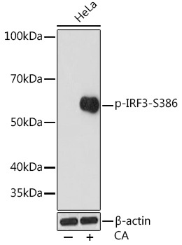
Western blot analysis of lysates from HeLa cells, using Phospho-IRF3-S386 Rabbit mAb (CABP0995) at 1:1000 dilution. HeLa cells were treated with Calyculin A (100 nM) at 37℃ for 30 minutes after serum-starvation overnight. Secondary antibody: HRP-conjugated Goat anti-Rabbit IgG (H+L) (CABS014) at 1:10000 dilution. Lysates/proteins: 25μg per lane. Blocking buffer: 3% BSA. Detection: ECL Enhanced Kit (AbGn00021). Exposure time: 3min.