The Phospho-JunD-S255 Monoclonal Antibody (CABP1142) is a high-quality antibody developed for reliable detection and analysis of target proteins. The protein encoded by this intronless gene is a member of the JUN family, and a functional component of the AP1 transcription factor complex. This protein has been proposed to protect cells from p53-dependent senescence and apoptosis. Alternative translation initiation site usage results in the production of different isoforms (PMID:12105216). RRID AB_2864009 Gene ID 3727 Swiss Prot Synonym AP-1; Phospho-JunD-S255
This antibody is validated for use in WB, ELISA applications and has demonstrated reactivity against Mouse samples.
Product Name:
Phospho-JunD-S255 Monoclonal Antibody
SKU:
CABP1142
Size:
100μL, 20μL
Reactivity:
Mouse
Clone Number:
ARC1603
Conjugate:
Unconjugated
Immunogen:
Synthetic peptide. This information is considered to be commercially sensitive.
Tested Applications:
WBELISA
Recommended Dilution:
WB
1:500 - 1:2000
ELISA
Recommended starting concentration is 1 μg/mL. Please optimize the concentration based on your specific assay requirements.
Synonyms:
AP-1, Phospho-JunD-S255
Positive Sample:
NIH/3T3 treated with Anisomycin
Cellular Localization:
Nucleus.
Calculated MW:
35kDa
Observed MW:
38kDa/42kDa
The protein encoded by this intronless gene is a member of the JUN family, and a functional component of the AP1 transcription factor complex. This protein has been proposed to protect cells from p53-dependent senescence and apoptosis. Alternative translation initiation site usage results in the production of different isoforms (PMID:12105216). RRID AB_2864009 Gene ID 3727 Swiss Prot Synonym AP-1; Phospho-JunD-S255
Purification Method:
Affinity purification
Gene ID:
3727
RRID:
AB_2864009
Buffer Information:
Store at -20℃. Avoid freeze / thaw cycles. Buffer: PBS containing 50% glycerol and 0.05% BSA, preserved with proclin300 or sodium azide, pH 7.3.
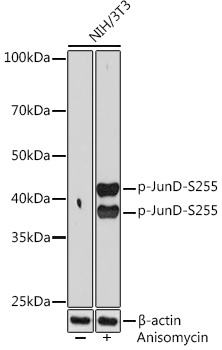
Western blot analysis of lysates from NIH/3T3 cells, using Phospho-JunD-S255 Rabbit mAb (CABP1142) at 1:1000 dilution. NIH/3T3 cells were treated with Anisomycin (25 μg/mL) at 37℃ for 30 minutes. Secondary antibody: HRP-conjugated Goat anti-Rabbit IgG (H+L) (AS014) at 1:10000 dilution. Lysates/proteins: 25μg per lane. Blocking buffer: 3% BSA. Detection: ECL Basic Kit (AbGn00020). Exposure time: 90s.