The Phospho-MARCKS-S162 Antibody (CABP0403) is a high-quality antibody developed for reliable detection and analysis of target proteins. This antibody, generated in rabbits, has high specificity for phosphorylated MARCKS at serine 162 in human samples, making it ideal for Western blot applications.MARCKS is a key player in cellular processes such as cell migration, adhesion, and neurite outgrowth, as well as in diseases like cancer and neurological disorders. The phosphorylation of MARCKS at serine 162 is known to impact its function, making this antibody essential for studying the role of MARCKS in cellular signaling and disease progression.
This antibody is validated for use in WB, ELISA applications and has demonstrated reactivity against Human, Mouse, Rat samples.
Product Name:
Phospho-MARCKS-S162 Antibody
SKU:
CABP0403
Size:
20μL, 100μL
Reactivity:
Human, Mouse, Rat
Conjugate:
Unconjugated
Immunogen:
Synthetic peptide. This information is considered to be commercially sensitive.
Tested Applications:
WBELISA
Recommended Dilution:
WB
1:100 - 1:500
ELISA
Recommended starting concentration is 1 μg/mL. Please optimize the concentration based on your specific assay requirements.
Synonyms:
MACS, 80K-L, PKCSL, PRKCSL, Phospho-MARCKS-S162
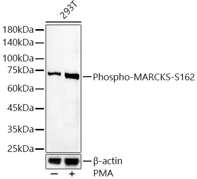
Positive Sample:
293T treated with PMA
Cellular Localization:
Cytoplasm, Lipid-Anchor, Membrane, Cytoskeleton.
Calculated MW:
32kDa
Observed MW:
75kDa
The protein encoded by this gene is a substrate for protein kinase C. It is localized to the plasma membrane and is an actin filament crosslinking protein. Phosphorylation by protein kinase C or binding to calcium-calmodulin inhibits its association with actin and with the plasma membrane, leading to its presence in the cytoplasm. The protein is thought to be involved in cell motility, phagocytosis, membrane trafficking and mitogenesis.
Purification Method
Affinity purification
Gene ID
4082
RRID
AB_2771321
Buffer Information
Store at -20℃. Avoid freeze / thaw cycles. Buffer: PBS containing 50% glycerol, preserved with proclin300 or sodium azide, pH 7.3.
Western blot analysis of lysates from 293T cells, using Phospho-MARCKS-S162 Rabbit pAb (CABP0403) at 1:400 dilution. 293T cells were treated with PMA/TPA (200 nM) at 37℃ for 30 minutes after serum-starvation overnight. Secondary antibody: HRP-conjugated Goat anti-Rabbit IgG (H+L) (CABS014) at 1:10000 dilution. Lysates/proteins: 25μg per lane. Blocking buffer: 3% nonfat dry milk in TBST. Detection: ECL Basic Kit (AbGn00020). Exposure time: 180s.