The Phospho-TBK1/NAK-S172 Monoclonal Antibody (CABP1026) is a high-quality antibody developed for reliable detection and analysis of target proteins. This antibody, developed using advanced monoclonal technology, specifically targets the phosphorylated form of NaK-TBK1 at serine 172. It is highly sensitive and selective, making it ideal for detecting and studying this important signaling pathway.NaK-TBK1 is a key regulator of innate immune responses and is involved in various cellular processes, including inflammation, autophagy, and antiviral responses. Phosphorylation of NaK-TBK1 at serine 172 is a crucial modification that activates its kinase activity, leading to downstream signaling cascades.
This antibody is validated for use in WB, ELISA applications and has demonstrated reactivity against Human, Mouse samples.
Product Name:
Phospho-TBK1/NAK-S172 Monoclonal Antibody
SKU:
CABP1026
Size:
20μL, 100μL
Reactivity:
Human, Mouse
Clone Number:
ARC1571
Conjugate:
Unconjugated
Immunogen:
Synthetic peptide. This information is considered to be commercially sensitive.
Sequence:
FVSL Y
Tested Applications:
WBELISA
Recommended Dilution:
WB
1:500 - 1:2000
ELISA
Recommended starting concentration is 1 μg/mL. Please optimize the concentration based on your specific assay requirements.
Synonyms:
NAK, T2K, IIAE8, FTDALS4, Phospho-TBK1/NAK-S172
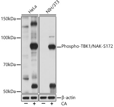
Positive Sample:
HeLa treated with Calyculin A, NIH/3T3 treated with Calyculin A
Cellular Localization:
Cytoplasm.
Calculated MW:
84kDa
Observed MW:
84kDa
The NF-kappa-B (NFKB) complex of proteins is inhibited by I-kappa-B (IKB) proteins, which inactivate NFKB by trapping it in the cytoplasm. Phosphorylation of serine residues on the IKB proteins by IKB kinases marks them for destruction via the ubiquitination pathway, thereby allowing activation and nuclear translocation of the NFKB complex. The protein encoded by this gene is similar to IKB kinases and can mediate NFKB activation in response to certain growth factors. The protein is also an important kinase for antiviral innate immunity response.
Purification Method
Affinity purification
Gene ID
29110
RRID
AB_2863910
Buffer Information
Store at -20℃. Avoid freeze / thaw cycles. Buffer: PBS containing 50% glycerol and 0.05% BSA, preserved with proclin300 or sodium azide, pH 7.3.
Western blot analysis of various lysates using Phospho-TBK1/NAK-S172 Rabbit mAb (CABP1026) at 1:1000 dilution. Both HeLa cells and NIH/3T3 cells were treated with Calyculin A (100 nM) at 37℃ for 30 minutes after serum-starvation overnight. Secondary antibody: HRP-conjugated Goat anti-Rabbit IgG (H+L) (CABS014) at 1:10000 dilution. Lysates/proteins: 25μg per lane. Blocking buffer: 3% BSA. Detection: ECL Basic Kit (AbGn00020). Exposure time: 1min.