The Phospho-NF-kB p65/RelA-T505 Polyclonal Antibody (CABP1318) is a high-quality antibody developed for reliable detection and analysis of target proteins. NF-kB p65 (RelA) is a key component of the NF-kB complex, which plays a critical role in regulating the expression of genes involved in immune response, inflammation, and cell survival.This antibody specifically targets the phosphorylated form of NF-kB p65 at threonine 505, a post-translational modification that regulates the activity of NF-kB in response to various stimuli. By detecting and quantifying phosphorylated NF-kB p65, researchers can gain insights into the activation status of the NF-kB pathway in different cell types and experimental conditions.
This antibody is validated for use in WB, ELISA applications and has demonstrated reactivity against Human samples.
Product Name:
Phospho-NF-kB p65/RelA-T505 Polyclonal Antibody
SKU:
CABP1318
Size:
20μL, 100μL
Reactivity:
Human
Conjugate:
Unconjugated
Immunogen:
Synthetic peptide. This information is considered to be commercially sensitive.
Sequence:
LVTG A
Tested Applications:
WBELISA
Recommended Dilution:
WB
1:500 - 1:2000
ELISA
Recommended starting concentration is 1 μg/mL. Please optimize the concentration based on your specific assay requirements.
NF-kappa-B is a ubiquitous transcription factor involved in several biological processes. It is held in the cytoplasm in an inactive state by specific inhibitors. Upon degradation of the inhibitor, NF-kappa-B moves to the nucleus and activates transcription of specific genes. NF-kappa-B is composed of NFKB1 or NFKB2 bound to either REL, RELA, or RELB. The most abundant form of NF-kappa-B is NFKB1 complexed with the product of this gene, RELA. Four transcript variants encoding different isoforms have been found for this gene.
Purification Method
Affinity purification
Gene ID
5970
Buffer Information
Store at -20℃. Avoid freeze / thaw cycles. Buffer: PBS containing 50% glycerol, preserved with proclin300 or sodium azide, pH 7.3.
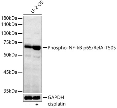
Western blot analysis of lysates from U-2 OS cells, using Phospho-NF-kB p65/RelA-T505 Rabbit pAb (CABP1318) at 1:1000 dilution. U-2 OS cells were treated with cisplatin at 37℃ for 16 hours. Secondary antibody: HRP-conjugated Goat anti-Rabbit IgG (H+L) (CABS014) at 1:10000 dilution. Lysates/proteins: 25μg per lane. Blocking buffer: 3% nonfat dry milk in TBST. Detection: ECL Basic Kit (AbGn00020). Exposure time: 90s.
 at 1:1000 dilution. U-2 OS cells were treated by cisplatin at 37℃ for 16 hours. Secondary antibody: HRP Goat Anti-Rabbit IgG (H+L) at 1:10000 dilution. Lysates/proteins: 25ug per lane. Blocking buffer: 3% nonfat dry milk in TBST.")
 at 1:1000 dilution. U-2 OS cells were treated by cisplatin at 37℃ for 16 hours. Secondary antibody: HRP Goat Anti-Rabbit IgG (H+L) at 1:10000 dilution. Lysates/proteins: 25ug per lane. Blocking buffer: 3% nonfat dry milk in TBST.")


