The Phospho-NPM1-T199 Antibody (CABP0836) is a high-quality antibody developed for reliable detection and analysis of target proteins. This antibody, produced in rabbits, exhibits high specificity and reactivity towards human samples, making it ideal for Western blot applications. By targeting the T199 phosphorylation site on NPM1, this antibody enables precise detection and analysis of NPM1 protein levels in different cell types.NPM1 plays a crucial role in regulating cell growth, proliferation, and apoptosis, making it a key player in cancer biology and other diseases.
This antibody is validated for use in WB, ELISA applications and has demonstrated reactivity against Human samples.
Product Name:
Phospho-NPM1-T199 Antibody
SKU:
CABP0836
Size:
20μL, 100μL
Reactivity:
Human
Conjugate:
Unconjugated
Immunogen:
Synthetic peptide. This information is considered to be commercially sensitive.
Sequence:
RDTP A
Tested Applications:
WBELISA
Recommended Dilution:
WB
1:500 - 1:1000
ELISA
Recommended starting concentration is 1 μg/mL. Please optimize the concentration based on your specific assay requirements.
Synonyms:
B23, NPM, Phospho-NPM1-T199
Positive Sample:
HeLa treated with nocodazole
Cellular Localization:
Cytoplasm, Nucleus, Centrosome, Cytoskeleton, Microtubule Organizing Center, Nucleolus, Nucleoplasm.
Calculated MW:
33kDa
Observed MW:
38kDa
The protein encoded by this gene is involved in several cellular processes, including centrosome duplication, protein chaperoning, and cell proliferation. The encoded phosphoprotein shuttles between the nucleolus, nucleus, and cytoplasm, chaperoning ribosomal proteins and core histones from the nucleus to the cytoplasm. This protein is also known to sequester the tumor suppressor ARF in the nucleolus, protecting it from degradation until it is needed. Mutations in this gene are associated with acute myeloid leukemia. Dozens of pseudogenes of this gene have been identified.
Purification Method
Affinity purification
Gene ID
4869
RRID
AB_2771374
Buffer Information
Store at -20℃. Avoid freeze / thaw cycles. Buffer: PBS containing 50% glycerol, preserved with proclin300 or sodium azide, pH 7.3.
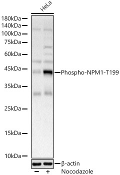
Western blot analysis of various lysates, using Phospho-NPM1-T199 Rabbit pAb (CABP0836) at 1:900 dilution. HeLa cells were treated with nocodazole (50 ng/ml) at 37℃ for 20 hours. Secondary antibody: HRP-conjugated Goat anti-Rabbit IgG (H+L) (CABS014) at 1:10000 dilution. Lysates/proteins: 25μg per lane. Blocking buffer: 3% nonfat dry milk in TBST. Detection: ECL Basic Kit (AbGn00020). Exposure time: 10s.