The Phospho-PAK2-S20 Antibody (CABP0803) is a high-quality antibody developed for reliable detection and analysis of target proteins. This antibody, generated in rabbits, is highly specific for human samples and has been validated for use in Western blot applications. It recognizes the phosphorylated form of Pak2 at serine 20, allowing for precise detection and analysis in a variety of cell types.Pak2 is a serine/threonine kinase that is involved in signaling pathways regulating cell growth and survival. Phosphorylation of Pak2 at serine 20 is known to enhance its kinase activity, influencing downstream cellular processes.
This antibody is validated for use in WB, ELISA applications and has demonstrated reactivity against Human, Mouse, Rat samples.
Product Name:
Phospho-PAK2-S20 Antibody
SKU:
CABP0803
Size:
20μL, 100μL
Reactivity:
Human, Mouse, Rat
Conjugate:
Unconjugated
Immunogen:
Synthetic peptide. This information is considered to be commercially sensitive.
Sequence:
MSST I
Tested Applications:
WBELISA
Recommended Dilution:
WB
1:500 - 1:2000
ELISA
Recommended starting concentration is 1 μg/mL. Please optimize the concentration based on your specific assay requirements.
Synonyms:
KNO2, PAK65, PAKgamma, Phospho-PAK2-S20
Positive Sample:
HeLa treated with Calyculin A, C6 treated with Calyculin A, NIH/3T3 treated with Calyculin A
The p21 activated kinases (PAK) are critical effectors that link Rho GTPases to cytoskeleton reorganization and nuclear signaling. The PAK proteins are a family of serine/threonine kinases that serve as targets for the small GTP binding proteins, CDC42 and RAC1, and have been implicated in a wide range of biological activities. The protein encoded by this gene is activated by proteolytic cleavage during caspase-mediated apoptosis, and may play a role in regulating the apoptotic events in the dying cell.
Purification Method
Affinity purification
Gene ID
5062
RRID
AB_2771401
Buffer Information
Store at -20℃. Avoid freeze / thaw cycles. Buffer: PBS containing 50% glycerol, preserved with proclin300 or sodium azide, pH 7.3.
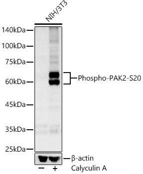
Western blot analysis of lysates from NIH/3T3 cells using Phospho-PAK2-S20 Rabbit pAb (CABP0803) at 1:1000 dilution. NIH/3T3 cells were treated with Calyculin A (100 nM) at 37℃ for 30 minutes after serum-starvation overnight. Secondary antibody: HRP-conjugated Goat anti-Rabbit IgG (H+L) (CABS014) at 1:10000 dilution. Lysates/proteins: 25 μg per lane. Blocking buffer: 3% nonfat dry milk in TBST. Detection: ECL Basic Kit (AbGn00020). Exposure time: 30s.
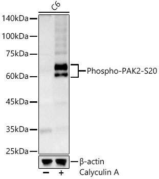
Western blot analysis of lysates from C6 cells using Phospho-PAK2-S20 Rabbit pAb (CABP0803) at 1:1000 dilution. C6 cells were treated with Calyculin A (100 nM) at 37℃ for 30 minutes after serum-starvation overnight. Secondary antibody: HRP-conjugated Goat anti-Rabbit IgG (H+L) (CABS014) at 1:10000 dilution. Lysates/proteins: 25 μg per lane. Blocking buffer: 3% nonfat dry milk in TBST. Detection: ECL Basic Kit (AbGn00020). Exposure time: 30s.