The Phospho-PAK4/PAK5/PAK6-S474/S560/S602 Monoclonal Antibody (CABP1049) is a high-quality antibody developed for reliable detection and analysis of target proteins. This antibody, generated in rabbits, exhibits high reactivity with human samples and is validated for use in Western blot applications.The PAK family of proteins are key regulators of cell proliferation, survival, and cytoskeletal organization, with dysregulation implicated in various diseases including cancer. Phosphorylation of PAK4, PAK5, and PAK6 at specific residues like S474, S560, and S602 has been shown to impact their functions, highlighting the importance of studying these modifications in cell signaling pathways.
This antibody is validated for use in WB, ELISA applications and has demonstrated reactivity against Mouse, Rat samples.
PAK proteins, a family of serine/threonine p21-activating kinases, include PAK1, PAK2, PAK3 and PAK4. PAK proteins are critical effectors that link Rho GTPases to cytoskeleton reorganization and nuclear signaling. They serve as targets for the small GTP binding proteins Cdc42 and Rac and have been implicated in a wide range of biological activities. PAK4 interacts specifically with the GTP-bound form of Cdc42Hs and weakly activates the JNK family of MAP kinases. PAK4 is a mediator of filopodia formation and may play a role in the reorganization of the actin cytoskeleton. Multiple alternatively spliced transcript variants encoding distinct isoforms have been found for this gene. [provided by RefSeq, Jul 2008]
Purification Method
Affinity purification
Gene ID
10298 57144 56924
RRID
AB_2863922
Buffer Information
Store at -20℃. Avoid freeze / thaw cycles. Buffer: PBS containing 50% glycerol and 0.05% BSA, preserved with proclin300 or sodium azide, pH 7.3.
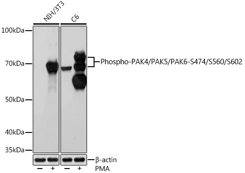
Western blot analysis of various lysates using Phospho-PAK4/PAK5/PAK6-S474/S560/S602 Rabbit mAb (CABP1049) at 1:1000 dilution. Both NIH/3T3 cells and C6 cells were treated with PMA/TPA (200 nM) at 37℃ for 30 minutes after serum-starvation overnight. Secondary antibody: HRP-conjugated Goat anti-Rabbit IgG (H+L) (CABS014) at 1:10000 dilution. Lysates/proteins: 25μg per lane. Blocking buffer: 3% BSA. Detection: ECL Basic Kit (AbGn00020). Exposure time: 10s.