The Phospho-PEA15-S104 Antibody (CABP0874) is a high-quality antibody developed for reliable detection and analysis of target proteins. PEA15, also known as phosphoprotein enriched in astrocytes 15 kDa, is a protein involved in cell survival, proliferation, and apoptosis pathways. This antibody, raised in rabbits, is highly specific for detecting phosphorylated PEA15 at serine 104 in human samples, making it an essential tool for Western blot applications.Phosphorylation of PEA15 at serine 104 has been implicated in various signaling pathways, including the MAPK/ERK pathway, which is crucial for cell growth and survival. Research has shown that dysregulation of PEA15 phosphorylation can lead to abnormal cell proliferation and contribute to the development of cancer and other diseases.
This antibody is validated for use in WB, IHC-P, ELISA applications and has demonstrated reactivity against Human, Mouse, Rat samples.
Product Name:
Phospho-PEA15-S104 Antibody
SKU:
CABP0874
Size:
20μL, 100μL
Reactivity:
Human, Mouse, Rat
Conjugate:
Unconjugated
Immunogen:
Synthetic peptide. This information is considered to be commercially sensitive.
Sequence:
IPSA K
Tested Applications:
WBIHC-PELISA
Recommended Dilution:
WB
1:500 - 1:2000
IHC-P
1:50 - 1:100
ELISA
Recommended starting concentration is 1 μg/mL. Please optimize the concentration based on your specific assay requirements.
This gene encodes a death effector domain-containing protein that functions as a negative regulator of apoptosis. The encoded protein is an endogenous substrate for protein kinase C. This protein is also overexpressed in type 2 diabetes mellitus, where it may contribute to insulin resistance in glucose uptake. Alternative splicing results in multiple transcript variants.
Purification Method
Affinity purification
Gene ID
8682
RRID
AB_2771412
Buffer Information
Store at -20℃. Avoid freeze / thaw cycles. Buffer: PBS with 0.01% thimerosal,50% glycerol,pH7.3.
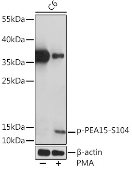
Western blot analysis of various lysates using Phospho-PEA15-S104 Rabbit pAb (CABP0874) at 1:1000 dilution. C6 cells were treated with PMA/TPA (200 nM) at 37℃ for 30 minutes after serum-starvation overnight. Secondary antibody: HRP-conjugated Goat anti-Rabbit IgG (H+L) (CABS014) at 1:10000 dilution. Lysates/proteins: 25μg per lane. Blocking buffer: 3% BSA. Detection: ECL Enhanced Kit (AbGn00021). Exposure time: 90s.