The Phospho-PERK-T982 Antibody (CABP0886) is a high-quality antibody developed for reliable detection and analysis of target proteins. The protein encoded by this gene phosphorylates the alpha subunit of eukaryotic translation-initiation factor 2, leading to its inactivation, and thus to a rapid reduction of translational initiation and repression of global protein synthesis. This protein is thought to modulate mitochondrial function. It is a type I membrane protein located in the endoplasmic reticulum (ER), where it is induced by ER stress caused by malfolded proteins. Mutations in this gene are associated with Wolcott-Rallison syndrome.
This antibody is validated for use in WB, IHC-P, ELISA applications and has demonstrated reactivity against Human, Mouse, Rat samples.
Product Name:
Phospho-PERK-T982 Antibody
SKU:
CABP0886
Size:
100μL, 20μL
Reactivity:
Human, Mouse, Rat
Conjugate:
Unconjugated
Immunogen:
Synthetic peptide. This information is considered to be commercially sensitive.
Tested Applications:
WBIHC-PELISA
Recommended Dilution:
WB
1:500 - 1:1000
IHC-P
1:50 - 1:200
ELISA
Recommended starting concentration is 1 μg/mL. Please optimize the concentration based on your specific assay requirements.
Synonyms:
PEK, WRS, PERK, Phospho-PERK-T982
Positive Sample:
293T treated with IGF-1, A549 treated with FBS
Cellular Localization:
Endoplasmic Reticulum Membrane, Single-Pass Type I Membrane Protein.
Calculated MW:
125kDa
Observed MW:
170kDa/
The protein encoded by this gene phosphorylates the alpha subunit of eukaryotic translation-initiation factor 2, leading to its inactivation, and thus to a rapid reduction of translational initiation and repression of global protein synthesis. This protein is thought to modulate mitochondrial function. It is a type I membrane protein located in the endoplasmic reticulum (ER), where it is induced by ER stress caused by malfolded proteins. Mutations in this gene are associated with Wolcott-Rallison syndrome.
Purification Method
Affinity purification
Gene ID
9451
RRID
AB_2771413
Buffer Information
Store at -20℃. Avoid freeze / thaw cycles. Buffer: PBS with 0.09% sodium azide,50% glycerol,pH7.3.
Western blot analysis of lysates from 293T cells, using Phospho-PERK-T982 Rabbit pAb (CABP0886) at 1:1000 dilution. 293T cells were treated with IGF-1 (50 ng/ml) at 37℃ for 5 minutes after serum-starvation overnight. Secondary antibody: HRP-conjugated Goat anti-Rabbit IgG (H+L) (AS014) at 1:10000 dilution. Lysates/proteins: 25μg per lane. Blocking buffer: 3% BSA. Detection: ECL Basic Kit (AbGn00020). Exposure time: 90s.
Western blot analysis of lysates from A549 cells using Phospho-PERK-T982 Rabbit pAb (CABP0886) at 1:1000 dilution incubated overnight at 4℃. A549 cells were treated with 10% FBS at 37℃ for 5 minutes after serum-starvation overnight. Secondary antibody: HRP-conjugated Goat anti-Rabbit IgG (H+L) (AS014) at 1:10000 dilution. Lysates/proteins: 30 μg per lane. Blocking buffer: 3 % nonfat dry milk in TBST. Detection: ECL Basic Kit (AbGn00020). Exposure time: 180s.
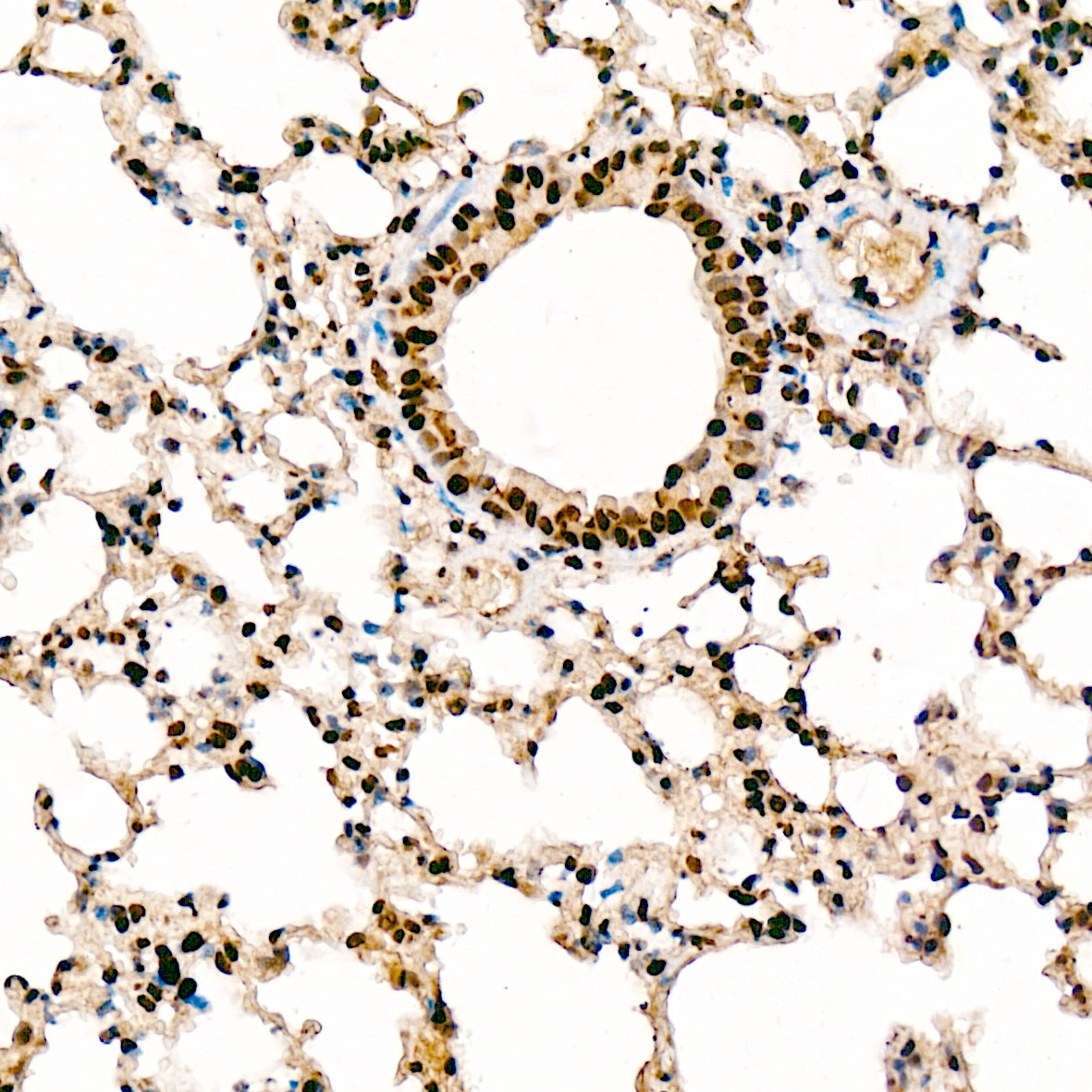
Immunohistochemistry analysis of paraffin-embedded Rat lung tissue using Phospho-PERK-T982 Rabbit pAb (CABP0886) at a dilution of 1:100 (40x lens). High pressure antigen retrieval was performed with 0.01 M citrate buffer (pH 6.0) prior to IHC staining.
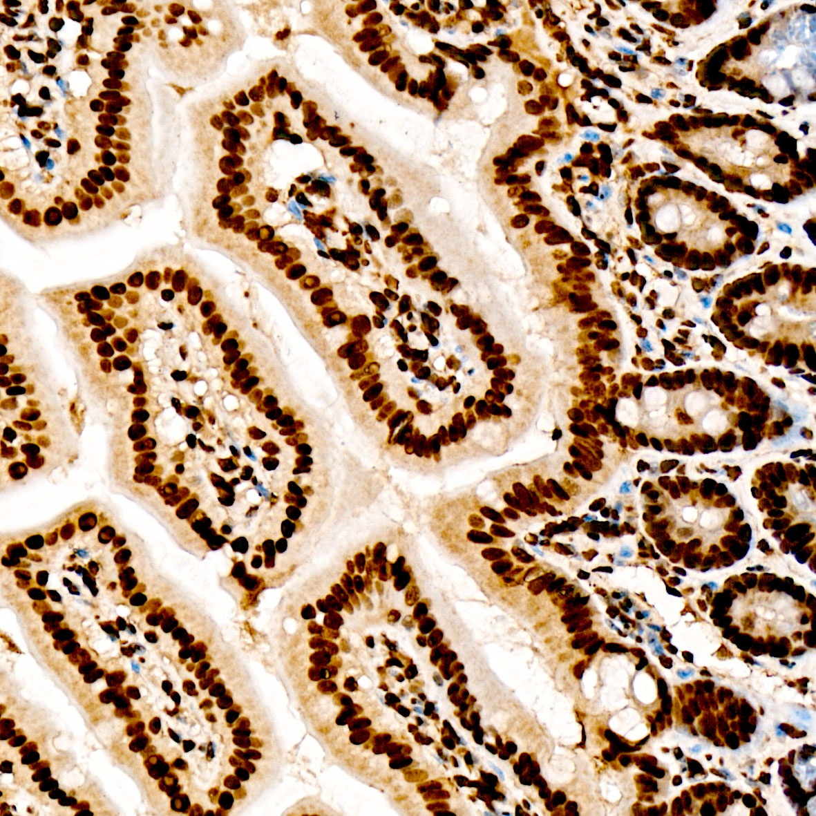
Immunohistochemistry analysis of paraffin-embedded Mouse Intestine tissue using Phospho-PERK-T982 Rabbit pAb (CABP0886) at a dilution of 1:100 (40x lens). High pressure antigen retrieval was performed with 0.01 M citrate buffer (pH 6.0) prior to IHC staining.