The PSMA3 Antibody (CAB1245) is a high-quality antibody developed for reliable detection and analysis of target proteins. This antibody is raised in rabbits and specifically targets PSMA3 in human samples, making it ideal for Western blot applications.PSMA3 plays a crucial role in maintaining cellular homeostasis by regulating protein turnover and degradation. Dysfunction in the proteasome complex, including PSMA3, has been implicated in various diseases, including cancer and neurodegenerative disorders.
This antibody is validated for use in WB, IHC-P, IP, ELISA, IF-P applications and has demonstrated reactivity against Human, Mouse, Rat samples.
Product Name:
PSMA3 Antibody
SKU:
CAB1245
Size:
20μL, 100μL
Reactivity:
Human, Mouse, Rat
Conjugate:
Unconjugated
Immunogen:
Recombinant protein (or fragment).This information is considered to be commercially sensitive.
0.5μg-4μg antibody for 200μg-400μg extracts of whole cells
IF-P
1:100 - 1:500
IHC-P
1:50 - 1:200
ELISA
Recommended starting concentration is 1 μg/mL. Please optimize the concentration based on your specific assay requirements.
Synonyms:
HC8, PSC3, PSMA3
Positive Sample:
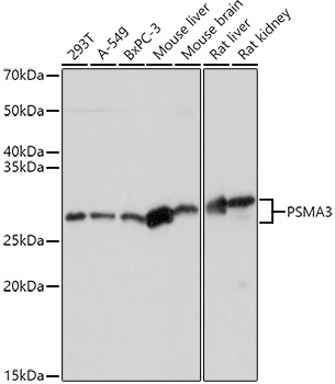
293T, A-549, BxPC-3, Mouse liver, Mouse brain, Rat liver, Rat kidney
Cellular Localization:
Cytoplasm, Nucleus.
Calculated MW:
28kDa
Observed MW:
28kDa
The proteasome is a multicatalytic proteinase complex with a highly ordered ring-shaped 20S core structure. The core structure is composed of 4 rings of 28 non-identical subunits; 2 rings are composed of 7 alpha subunits and 2 rings are composed of 7 beta subunits. Proteasomes are distributed throughout eukaryotic cells at a high concentration and cleave peptides in an ATP/ubiquitin-dependent process in a non-lysosomal pathway. An essential function of a modified proteasome, the immunoproteasome, is the processing of class I MHC peptides. This gene encodes a member of the peptidase T1A family, that is a 20S core alpha subunit. Two alternative transcripts encoding different isoforms have been identified.
Purification Method
Affinity purification
Gene ID
5684
RRID
AB_2759293
Buffer Information
Store at -20℃. Avoid freeze / thaw cycles. Buffer: PBS with 0.09% Sodium azide,50% glycerol,pH7.3.
Western blot analysis of various lysates using PSMA3 Rabbit pAb (CAB1245) at 1:1000 dilution. Secondary antibody: HRP-conjugated Goat anti-Rabbit IgG (H+L) (CABS014) at 1:10000 dilution. Lysates/proteins: 25μg per lane. Blocking buffer: 3% nonfat dry milk in TBST. Detection: ECL Basic Kit (AbGn00020). Exposure time: 3s.
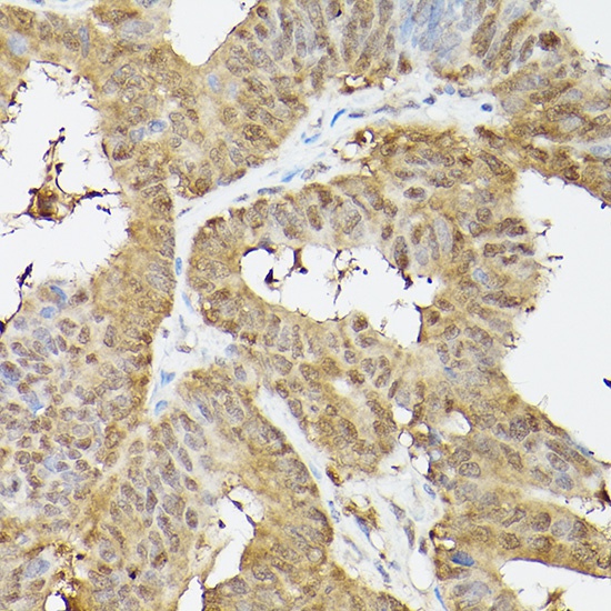
Immunohistochemistry analysis of paraffin-embedded Human colon carcinoma using PSMA3 Rabbit pAb (CAB1245) at dilution of 1:100 (40x lens). High pressure antigen retrieval performed with 0.01M Citrate buffer (pH 6.0) prior to IHC staining.
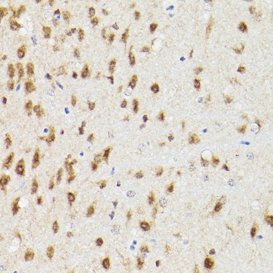
Immunohistochemistry analysis of paraffin-embedded Rat brain using PSMA3 Rabbit pAb (CAB1245) at dilution of 1:100 (40x lens). High pressure antigen retrieval performed with 0.01M Citrate buffer (pH 6.0) prior to IHC staining.
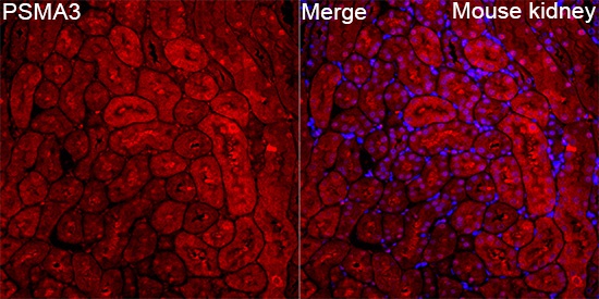
Immunofluorescence analysis of Mouse kidney tissue using PSMA3 Rabbit pAb (CAB1245) at a dilution of 1:400 (40x lens). Secondary antibody: Cy3-conjugated Goat anti-Rabbit IgG (H+L)(CABS007) at 1:500 dilution. Blue: DAPI for nuclear staining. High pressure antigen retrieval performed with 0.01M Citrate Buffer (pH 6.0) prior to IF staining.