The Phospho-NF-kB p65/RelA-S536 Antibody (CABP0475) is a high-quality antibody developed for reliable detection and analysis of target proteins. This antibody, raised in rabbits, specifically targets the phosphorylated form of RelA at serine 536, a key post-translational modification involved in the activation of NF-κB transcription factor activity.RelA, also known as p65, is a critical subunit of the NF-κB complex, which plays a central role in regulating immune responses, inflammation, cell survival, and proliferation. Phosphorylation of RelA at S536 is a key regulatory event that promotes its translocation into the nucleus and enhances its transcriptional activity in response to various stimuli, including pro-inflammatory cytokines and oxidative stress.
This antibody is validated for use in WB, IHC-P, ELISA applications and has demonstrated reactivity against Human, Mouse, Rat samples.
Product Name:
Phospho-NF-kB p65/RelA-S536 Antibody
SKU:
CABP0475
Size:
20μL, 100μL
Reactivity:
Human, Mouse, Rat
Conjugate:
Unconjugated
Immunogen:
Synthetic peptide. This information is considered to be commercially sensitive.
Sequence:
FSSI A
Tested Applications:
WBIHC-PELISA
Recommended Dilution:
WB
1:17000 - 1:71000
IHC-P
1:50 - 1:200
ELISA
Recommended starting concentration is 1 μg/mL. Please optimize the concentration based on your specific assay requirements.
NF-kappa-B is a ubiquitous transcription factor involved in several biological processes. It is held in the cytoplasm in an inactive state by specific inhibitors. Upon degradation of the inhibitor, NF-kappa-B moves to the nucleus and activates transcription of specific genes. NF-kappa-B is composed of NFKB1 or NFKB2 bound to either REL, RELA, or RELB. The most abundant form of NF-kappa-B is NFKB1 complexed with the product of this gene, RELA. Four transcript variants encoding different isoforms have been found for this gene.
Purification Method
Affinity purification
Gene ID
5970
RRID
AB_2771511
Buffer Information
Store at -20℃. Avoid freeze / thaw cycles. Buffer: PBS containing 50% glycerol, preserved with proclin300 or sodium azide, pH 7.3.
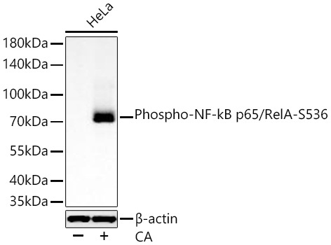
Western blot analysis of lysates from HeLa cells using Phospho-NF-kB p65/RelA-S536 Rabbit pAb (CABP0475) at 1:17000 dilution incubated overnight at 4℃. HeLa cells were treated with CA (100 nM) at 37℃ for 30 minutes after serum-starvation overnight. Secondary antibody: HRP-conjugated Goat anti-Rabbit IgG (H+L) (CABS014) at 1:10000 dilution. Lysates/proteins: 30 μg per lane. Blocking buffer: 3% nonfat dry milk in TBST. Detection: ECL Basic Kit (AbGn00020). Exposure time: 10 s.
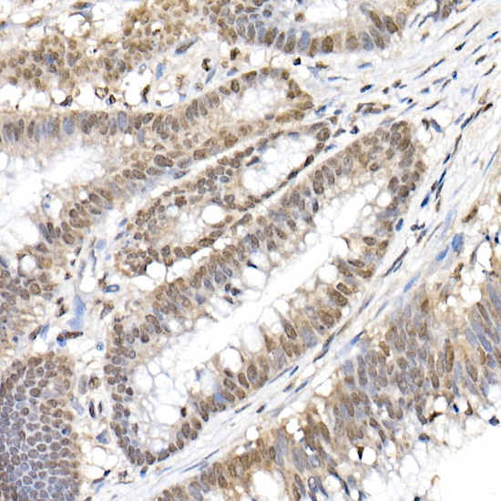
Immunohistochemistry analysis of paraffin-embedded Human colon carcinoma using Phospho-NF-kB p65/RelA-S536 Rabbit pAb (CABP0475) at dilution of 1:50 (40x lens). High pressure antigen retrieval performed with 0.01M Citrate buffer (pH 6.0) prior to IHC staining.