The Phospho-RPS6KA5-S376 Antibody (CABP0800) is a high-quality antibody developed for reliable detection and analysis of target proteins. This antibody, produced in rabbits, has been optimized for detection of phosphorylation at serine 376 in human samples, making it a valuable asset for studies on cell signaling, cancer biology, and drug development.RPS6KA5, also known as ribosomal protein S6 kinase A5, is a key player in regulating cell growth, proliferation, and differentiation through its phosphorylation of target proteins. Phosphorylation at serine 376 has been linked to specific cellular responses and may serve as a biomarker for various diseases and conditions.
This antibody is validated for use in WB, IHC-P, ELISA applications and has demonstrated reactivity against Human, Mouse, Rat samples.
Product Name:
Phospho-RPS6KA5-S376 Antibody
SKU:
CABP0800
Size:
20μL, 100μL
Reactivity:
Human, Mouse, Rat
Conjugate:
Unconjugated
Immunogen:
Synthetic peptide. This information is considered to be commercially sensitive.
Sequence:
GYSF V
Tested Applications:
WBIHC-PELISA
Recommended Dilution:
WB
1:500 - 1:2000
IHC-P
1:50 - 1:100
ELISA
Recommended starting concentration is 1 μg/mL. Please optimize the concentration based on your specific assay requirements.
Synonyms:
MSK1, RLPK, MSPK1, Phospho-RPS6KA5-S376
Positive Sample:
NIH/3T3 treated with Anisomycin
Cellular Localization:
Cytoplasm, Nucleus.
Calculated MW:
90kDa
Observed MW:
90kDa
Enables ATP binding activity and protein serine/threonine kinase activity. Involved in several processes, including histone-serine phosphorylation; positive regulation of histone modification; and regulation of transcription, DNA-templated. Located in cytoplasm and nucleoplasm.
Purification Method
Affinity purification
Gene ID
9252
RRID
AB_2771526
Buffer Information
Store at -20℃. Avoid freeze / thaw cycles. Buffer: PBS with 0.01% thimerosal,50% glycerol,pH7.3.
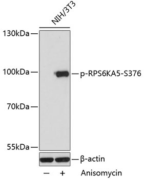
Western blot analysis of lysates from NIH/3T3 cells, using Phospho-RPS6KA5-S376 Rabbit pAb (CABP0800) at 1:2000 dilution. NIH/3T3 cells were treated with Anisomycin (25μg/mL) for 30 minutes. Secondary antibody: HRP-conjugated Goat anti-Rabbit IgG (H+L) (CABS014) at 1:10000 dilution. Lysates/proteins: 25μg per lane. Blocking buffer: 3% BSA. Detection: ECL Basic Kit (AbGn00020). Exposure time: 30s.
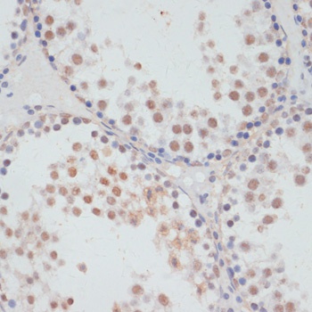
Immunohistochemistry analysis of paraffin-embedded Rat testis using Phospho-RPS6KA5-S376 Rabbit pAb (CABP0800) at dilution of 1:100 (40x lens). Microwave antigen retrieval performed with 0.01M Tris/EDTA Buffer (pH 9.0) prior to IHC staining.
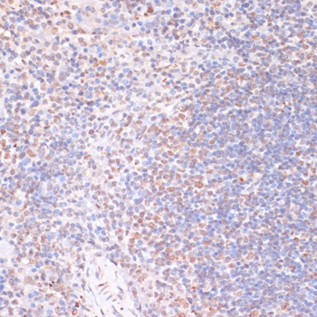
Immunohistochemistry analysis of paraffin-embedded Mouse spleen using Phospho-RPS6KA5-S376 Rabbit pAb (CABP0800) at dilution of 1:100 (40x lens). Microwave antigen retrieval performed with 0.01M Tris/EDTA Buffer (pH 9.0) prior to IHC staining.