The Phospho-Smad2-S465/S467 Monoclonal Antibody (CABP1342) is a high-quality antibody developed for reliable detection and analysis of target proteins. This antibody, produced in mice, specifically recognizes the phosphorylated form of Smad2 at residues S465 and S467, allowing for precise detection and quantification in Western blot experiments.Smad2 is a critical mediator of TGF-beta signaling, which is involved in a wide range of cellular processes, including cell growth, differentiation, and apoptosis. Phosphorylation at S465 and S467 is known to regulate the activity of Smad2, making this antibody essential for studying the functional implications of this post-translational modification.
This antibody is validated for use in WB, ELISA applications and has demonstrated reactivity against Human, Mouse samples.
Product Name:
Phospho-Smad2-S465/S467 Monoclonal Antibody
SKU:
CABP1342
Size:
20μL, 100μL
Reactivity:
Human, Mouse
Clone Number:
ARC56317
Conjugate:
Unconjugated
Immunogen:
Synthetic peptide. This information is considered to be commercially sensitive.
Sequence:
SSMS
Tested Applications:
WBELISA
Recommended Dilution:
WB
1:5000 - 1:20000
ELISA
Recommended starting concentration is 1 μg/mL. Please optimize the concentration based on your specific assay requirements.
The protein encoded by this gene belongs to the SMAD, a family of proteins similar to the gene products of the Drosophila gene 'mothers against decapentaplegic' (Mad) and the C. elegans gene Sma. SMAD proteins are signal transducers and transcriptional modulators that mediate multiple signaling pathways. This protein mediates the signal of the transforming growth factor (TGF)-beta, and thus regulates multiple cellular processes, such as cell proliferation, apoptosis, and differentiation. This protein is recruited to the TGF-beta receptors through its interaction with the SMAD anchor for receptor activation (SARA) protein. In response to TGF-beta signal, this protein is phosphorylated by the TGF-beta receptors. The phosphorylation induces the dissociation of this protein with SARA and the association with the family member SMAD4. The association with SMAD4 is important for the translocation of this protein into the nucleus, where it binds to target promoters and forms a transcription repressor complex with other cofactors. This protein can also be phosphorylated by activin type 1 receptor kinase, and mediates the signal from the activin. Alternatively spliced transcript variants have been observed for this gene.
Purification Method
Affinity purification
Gene ID
4087
Buffer Information
Store at -20℃. Avoid freeze / thaw cycles. Buffer: PBS containing 50% glycerol and 0.05% BSA, preserved with proclin300 or sodium azide, pH 7.3.
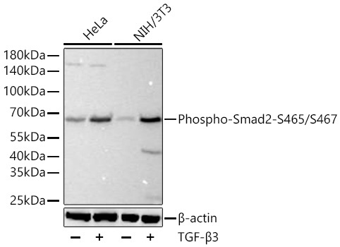
Western blot analysis of various lysates using Phospho-Smad2-S465/S467 Rabbit mAb (CABP1342) at 1:10000 dilution incubated at room temperature for 1.5 hours. HeLa cells and NIH/3T3 cells were treated with TGF-β3 (200 ng/mL) at 37℃ for 30 minutes after serum-starvation overnight. Secondary antibody: HRP-conjugated Goat anti-Rabbit IgG (H+L) (CABS014) at 1:10000 dilution. Lysates/proteins: 30 μg per lane. Blocking buffer: 3% nonfat dry milk in TBST. Detection: ECL Basic Kit (AbGn00020). Exposure time: 20s.
 at1:2000 dilution. HeLa and NIH/3T3 were treated by TGF-β3(H) (200 ng/mL) at 37℃ for 30 minutes after serum-starvation overnight. Secondary antibody: HRP Goat Anti-Rabbit IgG (H+L) at 1:10000 dilution. Lysates/proteins: 25μg per lane. Blocking buffer: 3% nonfat dry milk in TBST.")
 at1:2000 dilution. HeLa and NIH/3T3 were treated by TGF-β3(H) (200 ng/mL) at 37℃ for 30 minutes after serum-starvation overnight. Secondary antibody: HRP Goat Anti-Rabbit IgG (H+L) at 1:10000 dilution. Lysates/proteins: 25μg per lane. Blocking buffer: 3% nonfat dry milk in TBST.")


