The Phospho-Smad5-S463/S465 Polyclonal Antibody (CABP1319) is a high-quality antibody developed for reliable detection and analysis of target proteins. This antibody, generated in rabbits, specifically recognizes the phosphorylated form of Smad5 at serine residues 463 and 465. Validated for use in Western blot applications, this antibody allows for the detection and analysis of phosphorylated Smad5 in various cell types.The TGF-beta signaling pathway is essential for regulating cell growth, differentiation, and development, making the study of phosphorylated Smad5 crucial for understanding this important biological process.
This antibody is validated for use in WB, IHC-P, ELISA applications and has demonstrated reactivity against Human, Mouse, Rat samples.
Product Name:
Phospho-Smad5-S463/S465 Polyclonal Antibody
SKU:
CABP1319
Size:
20μL, 100μL
Reactivity:
Human, Mouse, Rat
Conjugate:
Unconjugated
Immunogen:
Synthetic peptide. This information is considered to be commercially sensitive.
Sequence:
SSVS
Tested Applications:
WBIHC-PELISA
Recommended Dilution:
WB
1:500 - 1:1000
IHC-P
1:50 - 1:200
ELISA
Recommended starting concentration is 1 μg/mL. Please optimize the concentration based on your specific assay requirements.
Synonyms:
DWFC, JV5-1, MADH5, Phospho-Smad5-S463/S465
Positive Sample:
NIH/3T3 treated with BMP4
Cellular Localization:
Cytoplasm, Cytosol, Nucleoplasm, Nucleus.
Calculated MW:
52kDa
The protein encoded by this gene is involved in the transforming growth factor beta signaling pathway that results in an inhibition of the proliferation of hematopoietic progenitor cells. The encoded protein is activated by bone morphogenetic proteins type 1 receptor kinase, and may be involved in cancer. Alternative splicing results in multiple transcript variants.
Purification Method
Affinity purification
Gene ID
4090
Buffer Information
Store at -20℃. Avoid freeze / thaw cycles. Buffer: PBS containing 50% glycerol, preserved with proclin300 or sodium azide, pH 7.3.
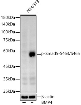
Western blot analysis of lysates from NIH/3T3 cells, using Phospho-Smad5-S463/S465 Rabbit pAb (CABP1319) at 1:600 dilution. NIH/3T3 cells were treated with BMP4 (50 ng/ml) at 37℃ for 30 minutes after serum-starvation overnight. Secondary antibody: HRP-conjugated Goat anti-Rabbit IgG (H+L) (CABS014) at 1:10000 dilution. Lysates/proteins: 25μg per lane. Blocking buffer: 3% nonfat dry milk in TBST. Detection: ECL Basic Kit (AbGn00020). Exposure time: 90s.
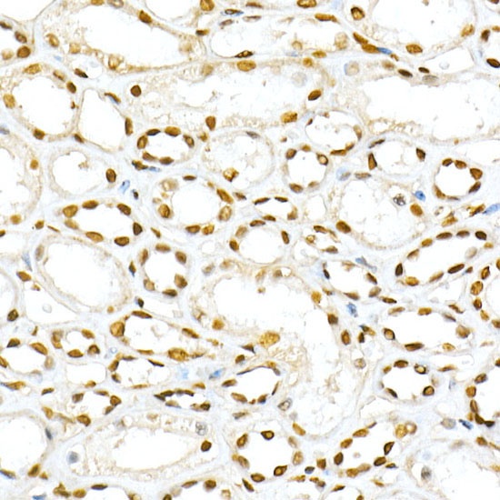
Immunohistochemistry analysis of paraffin-embedded Human kidney using Phospho-Smad5-S463/S465 Rabbit pAb (CABP1319) at dilution of 1:50 (40x lens). High pressure antigen retrieval performed with 0.01M Citrate buffer (pH 6.0) prior to IHC staining.
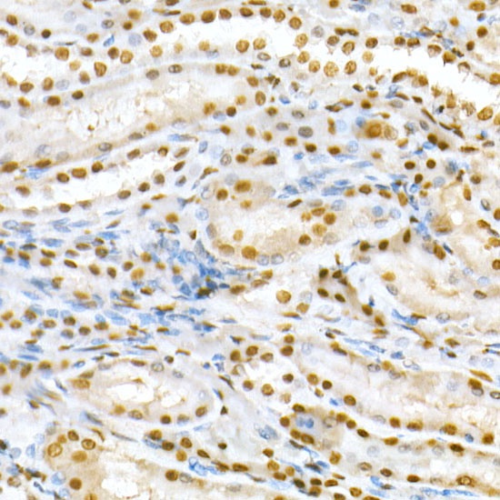
Immunohistochemistry analysis of paraffin-embedded Rat kidney using Phospho-Smad5-S463/S465 Rabbit pAb (CABP1319) at dilution of 1:50 (40x lens). High pressure antigen retrieval performed with 0.01M Citrate buffer (pH 6.0) prior to IHC staining.
 at 1:600 dilution. NIH/3T3 cells were treated by BMP4 (50 ng/ml) at 37℃ for 30 minutes after serum-starvation overnight. Secondary antibody: HRP Goat Anti-Rabbit IgG (H+L) at 1:10000 dilution. Lysates/proteins: 25μg per lane. Blocking buffer: 3% nonfat dry milk in TBST.")
 at 1:600 dilution. NIH/3T3 cells were treated by BMP4 (50 ng/ml) at 37℃ for 30 minutes after serum-starvation overnight. Secondary antibody: HRP Goat Anti-Rabbit IgG (H+L) at 1:10000 dilution. Lysates/proteins: 25μg per lane. Blocking buffer: 3% nonfat dry milk in TBST.")


