The Phospho-Smad5-S463/S465 Monoclonal Antibody (CABP1023) is a high-quality antibody developed for reliable detection and analysis of target proteins. This antibody, meticulously developed using rabbit monoclonal technology, offers high specificity and sensitivity when detecting phosphorylated Smad5 at serine residues 463 and 465 in human samples.Through its ability to bind specifically to phosphorylated Smad5, this antibody enables precise detection and quantification of Smad5 activation in various cell types and tissues. The phosphorylation of Smad5 at S463/S465 is known to play a crucial role in regulating cell growth, differentiation, and apoptosis, making this antibody an invaluable tool for researchers studying cellular signaling pathways, developmental biology, and cancer biology.
This antibody is validated for use in WB, ELISA applications and has demonstrated reactivity against Human samples.
Product Name:
Phospho-Smad5-S463/S465 Monoclonal Antibody
SKU:
CABP1023
Size:
20μL, 100μL
Reactivity:
Human
Clone Number:
ARC1568
Conjugate:
Unconjugated
Immunogen:
Synthetic peptide. This information is considered to be commercially sensitive.
Sequence:
SSVS
Tested Applications:
WBELISA
Recommended Dilution:
WB
1:500 - 1:1000
ELISA
Recommended starting concentration is 1 μg/mL. Please optimize the concentration based on your specific assay requirements.
Synonyms:
DWFC, JV5-1, MADH5, Phospho-Smad5-S463/S465
Positive Sample:
Hela treated with ATP
Cellular Localization:
Cytoplasm, Nucleus.
Calculated MW:
52kDa
Observed MW:
52kDa
The protein encoded by this gene is involved in the transforming growth factor beta signaling pathway that results in an inhibition of the proliferation of hematopoietic progenitor cells. The encoded protein is activated by bone morphogenetic proteins type 1 receptor kinase, and may be involved in cancer. Alternative splicing results in multiple transcript variants.
Purification Method
Affinity purification
Gene ID
4090
RRID
AB_2863908
Buffer Information
Store at -20℃. Avoid freeze / thaw cycles. Buffer: PBS containing 50% glycerol and 0.05% BSA, preserved with proclin300 or sodium azide, pH 7.3.
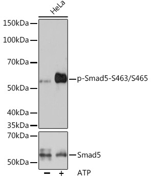
Western blot analysis of lysates from HeLa cells, using Phospho-Smad5-S463/S465 Rabbit mAb (CABP1023) at 1:1000 dilution or Smad5 antibody (CAB1947). Hela cells were treated with ATP(5 mM) at 30℃ for 1 hour. Secondary antibody: HRP-conjugated Goat anti-Rabbit IgG (H+L) (CABS014) at 1:10000 dilution. Lysates/proteins: 25μg per lane. Blocking buffer: 3% BSA. Detection: ECL Basic Kit (AbGn00020). Exposure time: 3min.