The Phospho-Smad5-S465 Monoclonal Antibody (CABP1411) is a high-quality antibody developed for reliable detection and analysis of target proteins. This antibody, produced using high quality monoclonal antibodies, specifically detects the phosphorylated form of Smad5 in human samples, making it an ideal choice for researchers studying signaling pathways involved in cell growth, differentiation, and development.The phosphorylation of Smad5 at serine 465 is known to play a crucial role in regulating the transcriptional activity of Smad proteins in response to TGF-beta signaling. By detecting and analyzing the phosphorylated form of Smad5, researchers can gain valuable insights into the regulatory mechanisms that control cell behavior and contribute to the development of diseases such as cancer, fibrosis, and developmental disorders.
This antibody is validated for use in WB, ELISA applications and has demonstrated reactivity against Human, Mouse, Rat samples.
Product Name:
Phospho-Smad5-S465 Monoclonal Antibody
SKU:
CABP1411
Size:
20μL, 100μL
Reactivity:
Human, Mouse, Rat
Clone Number:
ARC59204
Conjugate:
Unconjugated
Immunogen:
Synthetic peptide. This information is considered to be commercially sensitive.
Tested Applications:
WBELISA
Recommended Dilution:
WB
1:1000 - 1:5000
ELISA
Recommended starting concentration is 1 μg/mL. Please optimize the concentration based on your specific assay requirements.
Synonyms:
DWFC, JV5-1, MADH5, Phospho-Smad5-S465
Positive Sample:
NIH/3T3 treated with BMP4, C6 treated with TGFβ and MG132, HT-1080 treated with BMP2
Cellular Localization:
Cytoplasm, Nucleus.
Calculated MW:
52kDa
Observed MW:
52kDa/60kDa
The protein encoded by this gene is involved in the transforming growth factor beta signaling pathway that results in an inhibition of the proliferation of hematopoietic progenitor cells. The encoded protein is activated by bone morphogenetic proteins type 1 receptor kinase, and may be involved in cancer. Alternative splicing results in multiple transcript variants.
Purification Method
Affinity purification
Gene ID
4090
Buffer Information
Store at -20℃. Avoid freeze / thaw cycles. Buffer: PBS containing 50% glycerol and 0.05% BSA, preserved with proclin300 or sodium azide, pH 7.3.
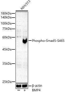
Western blot analysis of lysates from NIH/3T3 cells, using Phospho-Smad5-S465 Rabbit mAb (CABP1411) at 1:2000 dilution. NIH/3T3 cells were treated with BMP4(50ng/ml) at 37℃ for 6 hours. Secondary antibody: HRP-conjugated Goat anti-Rabbit IgG (H+L) (CABS014) at 1:10000 dilution. Lysates/proteins: 25μg per lane. Blocking buffer: 3% nonfat dry milk in TBST. Detection: ECL Basic Kit (AbGn00020). Exposure time: 180s.
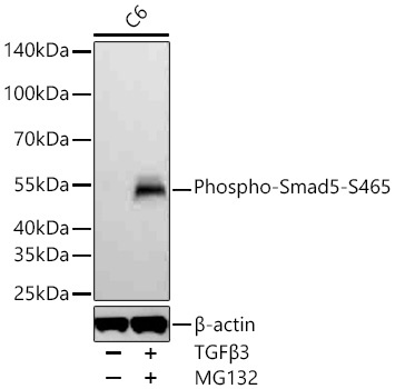
Western blot analysis of lysates from C6 cells using Phospho-Smad5-S465 Rabbit mAb (CABP1411) at 1:1000 dilution incubated overnight at 4℃. C6 cells were treated with TGFβ3 (10 ng/mL) and MG132 (10 μM) for 30 minutes after serum starvation. Secondary antibody: HRP-conjugated Goat anti-Rabbit IgG (H+L) (CABS014) at 1:10000 dilution. Lysates/proteins: 30 μg per lane. Blocking buffer: 3 % nonfat dry milk in TBST. Detection: ECL Basic Kit (AbGn00020). Exposure time: 20 s.
 at 1:2000 dilution. NIH/3T3 cells were treated by BMP4(50ng/ml) at 37℃ for 6 hours. Secondary antibody: HRP Goat Anti-Rabbit IgG (H+L) at 1:10000 dilution. Lysates/proteins: 25μg per lane. Blocking buffer: 3% nonfat dry milk in TBST.")
 at 1:2000 dilution. NIH/3T3 cells were treated by BMP4(50ng/ml) at 37℃ for 6 hours. Secondary antibody: HRP Goat Anti-Rabbit IgG (H+L) at 1:10000 dilution. Lysates/proteins: 25μg per lane. Blocking buffer: 3% nonfat dry milk in TBST.")


