The TPH1 Antibody (CAB1569) is a high-quality antibody developed for reliable detection and analysis of target proteins. This antibody, generated in rabbits, exhibits high specificity and reactivity with human samples, making it ideal for use in various research applications such as Western blotting. By binding to the TPH1 protein, this antibody enables accurate detection and analysis in different cell types, offering insights into serotonin regulation and its role in neurotransmission and gut function.TPH1, also known as tryptophan hydroxylase 1, is a key enzyme in the synthesis of serotonin, a neurotransmitter with diverse functions in the central nervous system and peripheral tissues.
This antibody is validated for use in WB, ELISA applications and has demonstrated reactivity against Human, Mouse, Rat samples.
Product Name:
TPH1 Antibody
SKU:
CAB1569
Size:
20μL, 100μL
Reactivity:
Human, Mouse, Rat
Conjugate:
Unconjugated
Immunogen:
Recombinant protein (or fragment).This information is considered to be commercially sensitive.
Recommended starting concentration is 1 μg/mL. Please optimize the concentration based on your specific assay requirements.
Synonyms:
TPRH, TRPH, TPH1
Positive Sample:
293F transfected with TPH1, Mouse brain, Rat colon
Cellular Localization:
Cytosol, Neuron Projection.
Calculated MW:
51kDa
Observed MW:
51kDa
This gene encodes a member of the aromatic amino acid hydroxylase family. The encoded protein catalyzes the first and rate limiting step in the biosynthesis of serotonin, an important hormone and neurotransmitter. Mutations in this gene have been associated with an elevated risk for a variety of diseases and disorders, including schizophrenia, somatic anxiety, anger-related traits, bipolar disorder, suicidal behavior, addictions, and others.
Purification Method
Affinity purification
Gene ID
7166
RRID
AB_2763100
Buffer Information
Store at -20℃. Avoid freeze / thaw cycles. Buffer: PBS containing 50% glycerol, preserved with proclin300 or sodium azide, pH 7.3.
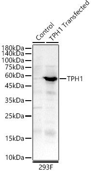
Western blot analysis of lysates from wild type (WT) and 293F cells transfected with TPH1 using TPH1 Rabbit pAb (CAB1569) at 1:1000 dilution. Secondary antibody: HRP-conjugated Goat anti-Rabbit IgG (H+L) (CABS014) at 1:10000 dilution. Lysates/proteins: 25μg per lane. Blocking buffer: 3% nonfat dry milk in TBST. Detection: ECL Basic Kit (AbGn00020). Exposure time: 90 s.
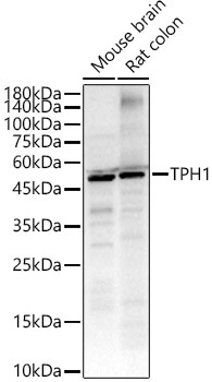
Western blot analysis of various lysates, using TPH1 Rabbit pAb (CAB1569) at 1:1000 dilution. Secondary antibody: HRP-conjugated Goat anti-Rabbit IgG (H+L) (CABS014) at 1:10000 dilution. Lysates/proteins: 25μg per lane. Blocking buffer: 3% nonfat dry milk in TBST. Detection: ECL Basic Kit (AbGn00020). Exposure time: 90s.