The PLA2G1B Antibody (CAB5478) is a high-quality antibody developed for reliable detection and analysis of target proteins. This antibody, generated in rabbits, exhibits high specificity and sensitivity towards human samples, making it ideal for use in Western blot applications.Phospholipase A2 plays a crucial role in various cellular processes, including inflammation, cell signaling, and membrane remodeling. Dysregulation of phospholipase A2 is implicated in various diseases, such as cardiovascular disorders, neurodegenerative diseases, and inflammatory conditions.
This antibody is validated for use in WB, IHC-P, ELISA applications and has demonstrated reactivity against Human, Mouse samples.
Product Name:
PLA2G1B Antibody
SKU:
CAB5478
Size:
20μL, 100μL
Reactivity:
Human, Mouse
Conjugate:
Unconjugated
Immunogen:
Recombinant protein (or fragment).This information is considered to be commercially sensitive.
Recommended starting concentration is 1 μg/mL. Please optimize the concentration based on your specific assay requirements.
Synonyms:
PLA2, PLA2A, PPLA2, PLA2G1B
Positive Sample:
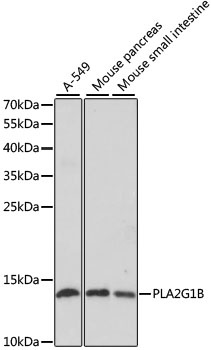
A-549, Mouse pancreas, Mouse small intestine
Cellular Localization:
Secreted.
Calculated MW:
16kDa
Observed MW:
16kDa
This gene encodes a secreted member of the phospholipase A2 (PLA2) class of enzymes, which is produced by the pancreatic acinar cells. The encoded calcium-dependent enzyme catalyzes the hydrolysis of the sn-2 position of membrane glycerophospholipids to release arachidonic acid (AA) and lysophospholipids. AA is subsequently converted by downstream metabolic enzymes to several bioactive lipophilic compounds (eicosanoids), including prostaglandins (PGs) and leukotrienes (LTs). The enzyme may be involved in several physiological processes including cell contraction, cell proliferation and pathological response.
Purification Method
Affinity purification
Gene ID
5319
RRID
AB_2766279
Buffer Information
Store at -20℃. Avoid freeze / thaw cycles. Buffer: PBS with 0.01% thimerosal,50% glycerol,pH7.3.
Western blot analysis of various lysates using PLA2G1B Rabbit pAb (CAB5478) at 1:1000 dilution. Secondary antibody: HRP-conjugated Goat anti-Rabbit IgG (H+L) (CABS014) at 1:10000 dilution. Lysates/proteins: 25μg per lane. Blocking buffer: 3% nonfat dry milk in TBST. Detection: ECL Enhanced Kit (AbGn00021). Exposure time: 90s.
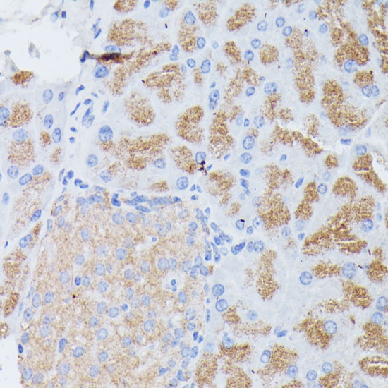
Immunohistochemistry analysis of paraffin-embedded Mouse pancreas using PLA2G1B Rabbit pAb (CAB5478) at dilution of 1:100 (40x lens). Microwave antigen retrieval performed with 0.01M PBS Buffer (pH 7.2) prior to IHC staining.