The PIGM Antibody (CAB17811) is a high-quality antibody developed for reliable detection and analysis of target proteins. This antibody, raised in rabbits, is highly specific for human samples and has been validated for use in Western blot applications.PIGM plays a critical role in anchoring proteins to the cell membrane, and mutations in the PIGM gene have been associated with glycosylphosphatidylinositol biosynthesis defect 7, a rare genetic disorder.
This antibody is validated for use in WB, IHC-P, IF/ICC, ELISA applications and has demonstrated reactivity against Human, Mouse, Rat samples.
Product Name:
PIGM Antibody
SKU:
CAB17811
Size:
20μL, 100μL
Reactivity:
Human, Mouse, Rat
Conjugate:
Unconjugated
Immunogen:
Recombinant protein (or fragment).This information is considered to be commercially sensitive.
Recommended starting concentration is 1 μg/mL. Please optimize the concentration based on your specific assay requirements.
Synonyms:
GPI-MT-I, PIGM
Positive Sample:
BxPC-3, Mouse thymus, Mouse spleen, Mouse lung, Rat thymus, Rat spleen
Calculated MW:
49kDa
Observed MW:
49kDa
This gene encodes a transmembrane protein that is located in the endoplasmic reticulum and is involved in GPI-anchor biosynthesis. The glycosylphosphatidylinositol (GPI)-anchor is a glycolipid which contains three mannose molecules in its core backbone. The GPI-anchor is found on many blood cells and serves to anchor proteins to the cell surface. This gene encodes a mannosyltransferase, GPI-MT-I, that transfers the first mannose to GPI on the lumenal side of the endoplasmic reticulum.
Purification Method
Affinity purification
Gene ID
93183
RRID
AB_2771657
Buffer Information
Store at -20℃. Avoid freeze / thaw cycles. Buffer: PBS with 0.01% thimerosal,50% glycerol,pH7.3.
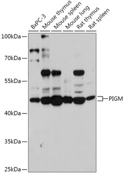
Western blot analysis of various lysates using PIGM Rabbit pAb (CAB17811) at 1:1000 dilution. Secondary antibody: HRP-conjugated Goat anti-Rabbit IgG (H+L) (CABS014) at 1:10000 dilution. Lysates/proteins: 25μg per lane. Blocking buffer: 3% nonfat dry milk in TBST. Detection: ECL Basic Kit (AbGn00020). Exposure time: 120s.
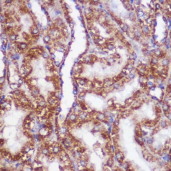
Immunohistochemistry analysis of paraffin-embedded Rat kidney using PIGM Rabbit pAb (CAB17811) at dilution of 1:100 (40x lens). Microwave antigen retrieval performed with 0.01M PBS Buffer (pH 7.2) prior to IHC staining.
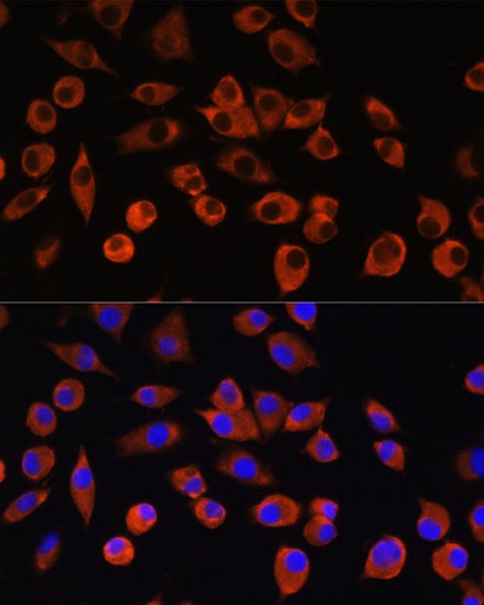
Immunofluorescence analysis of L929 cells using PIGM Rabbit pAb (CAB17811) at dilution of 1:100. Secondary antibody: Cy3-conjugated Goat anti-Rabbit IgG (H+L) (CABS007) at 1:500 dilution. Blue: DAPI for nuclear staining.