The PIK3R4/VPS15 Antibody (CAB15828) is a high-quality antibody developed for reliable detection and analysis of target proteins. Predicted to enable protein serine/threonine kinase activity. Involved in positive regulation of phosphatidylinositol 3-kinase activity; receptor catabolic process; and regulation of cytokinesis. Located in late endosome and microtubule cytoskeleton. RRID AB_2763252 Gene ID 30849 Swiss Prot Synonym p150; VPS15; PIK3R4/VPS15
This antibody is validated for use in WB, IHC-P, IF/ICC, ELISA applications and has demonstrated reactivity against Human, Mouse, Rat samples.
Product Name:
PIK3R4/VPS15 Antibody
SKU:
CAB15828
Size:
100μL, 20μL
Reactivity:
Human, Mouse, Rat
Clone Number:
-
Conjugate:
Unconjugated
Immunogen:
Recombinant protein (or fragment).This information is considered to be commercially sensitive.
Tested Applications:
WBIHC-PIF/ICCELISA
Recommended Dilution:
WB
1:500 - 1:1000
IHC-P
1:50 - 1:100
IF
/
ICC
1:50 - 1:200
ELISA
Recommended starting concentration is 1 μg/mL. Please optimize the concentration based on your specific assay requirements.
Synonyms:
p150, VPS15, PIK3R4/VPS15
Positive Sample:
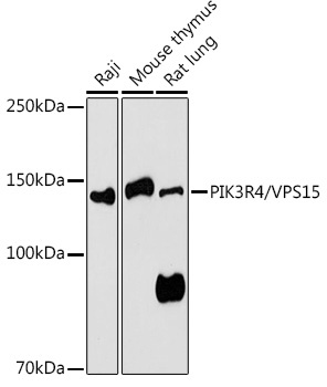
Raji, Mouse thymus, Rat lung
Cellular Localization:
Cytoplasmic Vesicle, Late Endosome, Lipid-Anchor, Membrane, Autophagosome.
Calculated MW:
153kDa
Observed MW:
150kDa
Predicted to enable protein serine/threonine kinase activity. Involved in positive regulation of phosphatidylinositol 3-kinase activity; receptor catabolic process; and regulation of cytokinesis. Located in late endosome and microtubule cytoskeleton. RRID AB_2763252 Gene ID 30849 Swiss Prot Synonym p150; VPS15; PIK3R4/VPS15
Purification Method:
Affinity purification
Gene ID:
30849
RRID:
AB_2763252
Buffer Information:
Store at -20℃. Avoid freeze / thaw cycles. Buffer: PBS containing 50% glycerol, preserved with proclin300 or sodium azide, pH 7.3.
Western blot analysis of various lysates using PIK3R4/VPS15 Rabbit pAb (CAB15828) at 1:1000 dilution. Secondary antibody: HRP-conjugated Goat anti-Rabbit IgG (H+L) (AS014) at 1:10000 dilution. Lysates/proteins: 25μg per lane. Blocking buffer: 3% nonfat dry milk in TBST. Detection: ECL Basic Kit (AbGn00020). Exposure time: 180s.
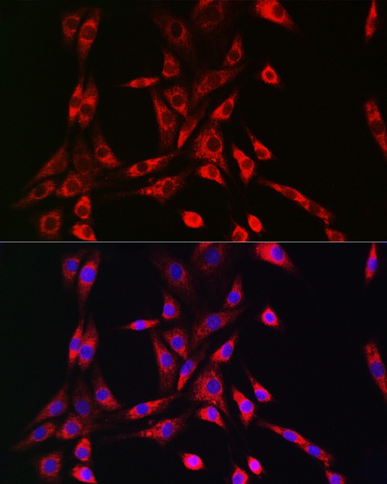
Immunofluorescence analysis of NIH/3T3 cells using PIK3R4/VPS15 Rabbit pAb (CAB15828) at dilution of 1:50 (40x lens). Secondary antibody: Cy3-conjugated Goat anti-Rabbit IgG (H+L) (AS007) at 1:500 dilution. Blue: DAPI for nuclear staining.