The PIM1 Monoclonal Antibody (CAB19695) is a high-quality antibody developed for reliable detection and analysis of target proteins. This antibody, generated in rabbits, exhibits high reactivity with human samples and has been validated for use in Western blot and immunohistochemistry applications.PIM1 is a critical player in signaling pathways that promote cell proliferation and inhibit apoptosis, making it a potential target for cancer therapy. Its aberrant expression has been linked to various types of cancer, making the PIM1 antibody an essential tool for studying the role of this kinase in oncogenesis.
This antibody is validated for use in WB, IHC-P, ELISA applications and has demonstrated reactivity against Human, Mouse, Rat samples.
Product Name:
PIM1 Monoclonal Antibody
SKU:
CAB19695
Size:
20μL, 100μL
Reactivity:
Human, Mouse, Rat
Clone Number:
ARC0175
Conjugate:
Unconjugated
Immunogen:
Synthetic peptide. This information is considered to be commercially sensitive.
Recommended starting concentration is 1 μg/mL. Please optimize the concentration based on your specific assay requirements.
Synonyms:
PIM, PIM1
Positive Sample:
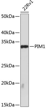
22Rv1
Cellular Localization:
Cell Membrane, Cytoplasm, Nucleus.
Calculated MW:
36kDa
Observed MW:
34kDa
The protein encoded by this gene belongs to the Ser/Thr protein kinase family, and PIM subfamily. This gene is expressed primarily in B-lymphoid and myeloid cell lines, and is overexpressed in hematopoietic malignancies and in prostate cancer. It plays a role in signal transduction in blood cells, contributing to both cell proliferation and survival, and thus provides a selective advantage in tumorigenesis. Both the human and orthologous mouse genes have been reported to encode two isoforms (with preferential cellular localization) resulting from the use of alternative in-frame translation initiation codons, the upstream non-AUG (CUG) and downstream AUG codons (PMIDs:16186805, 1825810).
Purification Method
Affinity purification
Gene ID
5292
RRID
AB_2862740
Buffer Information
Store at -20℃. Avoid freeze / thaw cycles. Buffer: PBS containing 50% glycerol and 0.05% BSA, preserved with proclin300 or sodium azide, pH 7.3.
Western blot analysis of lysates from 22Rv1 cells, using PIM1 Rabbit mAb (CAB19695) at 1:1000 dilution. Secondary antibody: HRP-conjugated Goat anti-Rabbit IgG (H+L) (CABS014) at 1:10000 dilution. Lysates/proteins: 25μg per lane. Blocking buffer: 3% nonfat dry milk in TBST. Detection: ECL Basic Kit (AbGn00020). Exposure time: 90s.
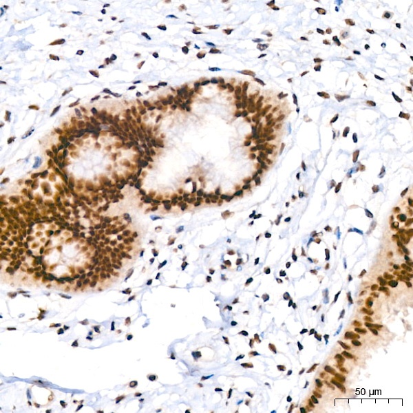
Immunohistochemistry analysis of paraffin-embedded Human cervix using PIM1 Rabbit mAb (CAB19695) at dilution of 1:200 (40x lens). High pressure antigen retrieval performed with 0.01M Tris/EDTA Buffer (pH 9.0) prior to IHC staining.
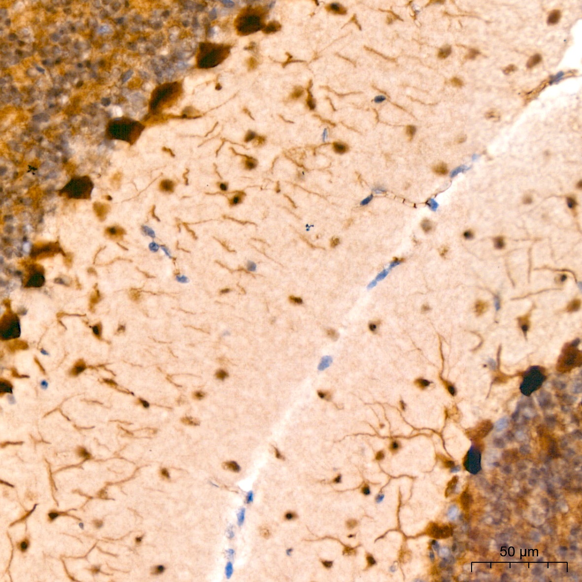
Immunohistochemistry analysis of paraffin-embedded Mouse brain using PIM1 Rabbit mAb (CAB19695) at dilution of 1:200 (40x lens). High pressure antigen retrieval performed with 0.01M Tris/EDTA Buffer (pH 9.0) prior to IHC staining.
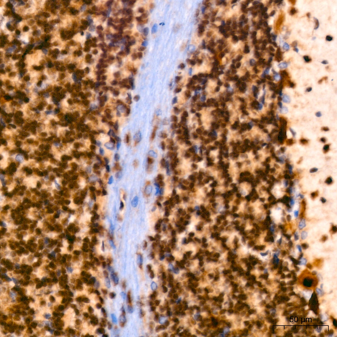
Immunohistochemistry analysis of paraffin-embedded Rat brain using PIM1 Rabbit mAb (CAB19695) at dilution of 1:200 (40x lens). High pressure antigen retrieval performed with 0.01M Tris/EDTA Buffer (pH 9.0) prior to IHC staining.