The PKD1L2 Antibody (CAB14954) is a high-quality antibody developed for reliable detection and analysis of target proteins. This gene encodes a member of the polycystin protein family. This protein may function as a G-protein-coupled component or regulator of cation channel pores. The long isoform of this protein contains 11 transmembrane domains, a latrophilin/CL-1-like GPCR proteolytic site (GPS) domain, and a polycystin-1, lipoxygenase, alpha-toxin (PLAT) domain. Alternative splicing results in multiple transcript variants encoding distinct isoforms. This gene is a polymorphic pseudogene in humans.
This antibody is validated for use in WB, ELISA applications and has demonstrated reactivity against Mouse samples.
Product Name:
PKD1L2 Antibody
SKU:
CAB14954
Size:
100μL, 20μL
Reactivity:
Mouse
Conjugate:
Unconjugated
Immunogen:
Recombinant protein (or fragment).This information is considered to be commercially sensitive.
Tested Applications:
WBELISA
Recommended Dilution:
WB
1:500 - 1:2000
ELISA
Recommended starting concentration is 1 μg/mL. Please optimize the concentration based on your specific assay requirements.
Synonyms:
PC1L2, PKD1L2
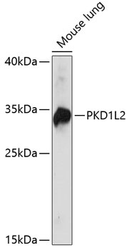
Positive Sample:
Mouse lung
Cellular Localization:
Membrane, Multi-Pass Membrane Protein.
Calculated MW:
273kDa
Observed MW:
33kDa
This gene encodes a member of the polycystin protein family. This protein may function as a G-protein-coupled component or regulator of cation channel pores. The long isoform of this protein contains 11 transmembrane domains, a latrophilin/CL-1-like GPCR proteolytic site (GPS) domain, and a polycystin-1, lipoxygenase, alpha-toxin (PLAT) domain. Alternative splicing results in multiple transcript variants encoding distinct isoforms. This gene is a polymorphic pseudogene in humans.
Purification Method
Affinity purification
Gene ID
114780
RRID
AB_2761837
Buffer Information
Store at -20℃. Avoid freeze / thaw cycles. Buffer: PBS with 0.01% thimerosal,50% glycerol,pH7.3.
Western blot analysis of lysates from mouse lung, using PKD1L2 Rabbit pAb (CAB14954) at 1:1000 dilution. Secondary antibody: HRP-conjugated Goat anti-Rabbit IgG (H+L) (AS014) at 1:10000 dilution. Lysates/proteins: 25μg per lane. Blocking buffer: 3% nonfat dry milk in TBST. Detection: ECL Basic Kit (AbGn00020). Exposure time: 30s.