The PKD1L2 Antibody (CAB14954) is a high-quality antibody developed for reliable detection and analysis of target proteins. This antibody, produced in rabbits, exhibits high specificity and sensitivity for human samples, making it suitable for Western blot analysis. By selectively binding to PKD1L2, the antibody facilitates accurate detection and analysis of the protein in different cell types, thereby facilitating investigations in the fields of cell biology and cancer research.PKD1L2 is known to play a crucial role in cell signaling pathways and the regulation of gene expression, making it a promising target for the development of therapeutic interventions.
This antibody is validated for use in WB, ELISA applications and has demonstrated reactivity against Mouse samples.
Product Name:
PKD1L2 Antibody
SKU:
CAB14954
Size:
20μL, 100μL
Reactivity:
Mouse
Conjugate:
Unconjugated
Immunogen:
Recombinant protein (or fragment).This information is considered to be commercially sensitive.
Recommended starting concentration is 1 μg/mL. Please optimize the concentration based on your specific assay requirements.
Synonyms:
PC1L2, PKD1L2
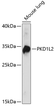
Positive Sample:
Mouse lung
Cellular Localization:
Membrane, Multi-Pass Membrane Protein.
Calculated MW:
273kDa
Observed MW:
33kDa
This gene encodes a member of the polycystin protein family. This protein may function as a G-protein-coupled component or regulator of cation channel pores. The long isoform of this protein contains 11 transmembrane domains, a latrophilin/CL-1-like GPCR proteolytic site (GPS) domain, and a polycystin-1, lipoxygenase, alpha-toxin (PLAT) domain. Alternative splicing results in multiple transcript variants encoding distinct isoforms. This gene is a polymorphic pseudogene in humans.
Purification Method
Affinity purification
Gene ID
114780
RRID
AB_2761837
Buffer Information
Store at -20℃. Avoid freeze / thaw cycles. Buffer: PBS with 0.01% thimerosal,50% glycerol,pH7.3.
Western blot analysis of lysates from mouse lung, using PKD1L2 Rabbit pAb (CAB14954) at 1:1000 dilution. Secondary antibody: HRP-conjugated Goat anti-Rabbit IgG (H+L) (CABS014) at 1:10000 dilution. Lysates/proteins: 25μg per lane. Blocking buffer: 3% nonfat dry milk in TBST. Detection: ECL Basic Kit (AbGn00020). Exposure time: 30s.