The PKNOX1 Antibody (CAB15301) is a high-quality antibody developed for reliable detection and analysis of target proteins. This antibody, produced in rabbits, is highly specific to human samples and is validated for use in various applications, including Western blotting.PKNOX1, also known as Prep1, is a transcription factor that plays a crucial role in cell differentiation and patterning during embryonic development. It is also implicated in regulating gene expression in various tissues, making it a key player in developmental biology and cancer research.
This antibody is validated for use in WB, ELISA applications and has demonstrated reactivity against Human, Mouse samples.
Product Name:
PKNOX1 Antibody
SKU:
CAB15301
Size:
20μL, 100μL
Reactivity:
Human, Mouse
Conjugate:
Unconjugated
Immunogen:
Recombinant protein (or fragment).This information is considered to be commercially sensitive.
Recommended starting concentration is 1 μg/mL. Please optimize the concentration based on your specific assay requirements.
Synonyms:
PREP1, pkonx1c, PKNOX1
Positive Sample:
293T, Mouse thymus
Cellular Localization:
Nucleus.
Calculated MW:
48kDa
Observed MW:
55kDa
Enables sequence-specific double-stranded DNA binding activity. Predicted to be involved in angiogenesis and regulation of transcription by RNA polymerase II. Predicted to act upstream of or within camera-type eye development; hemopoiesis; and positive regulation of transcription by RNA polymerase II. Predicted to be located in cytoplasm and nucleus. Predicted to be part of chromatin.
Purification Method
Affinity purification
Gene ID
5316
RRID
AB_2762203
Buffer Information
Store at -20℃. Avoid freeze / thaw cycles. Buffer: PBS with 0.01% thimerosal,50% glycerol,pH7.3.
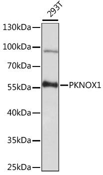
Western blot analysis of lysates from 293T cells, using PKNOX1 Rabbit pAb (CAB15301) at 1:1000 dilution. Secondary antibody: HRP-conjugated Goat anti-Rabbit IgG (H+L) (CABS014) at 1:10000 dilution. Lysates/proteins: 25μg per lane. Blocking buffer: 3% nonfat dry milk in TBST. Detection: ECL Basic Kit (AbGn00020). Exposure time: 30s.
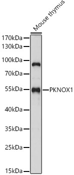
Western blot analysis of lysates from Mouse thymus, using PKNOX1 Rabbit pAb (CAB15301) at 1:1000 dilution. Secondary antibody: HRP-conjugated Goat anti-Rabbit IgG (H+L) (CABS014) at 1:10000 dilution. Lysates/proteins: 25μg per lane. Blocking buffer: 3% nonfat dry milk in TBST. Detection: ECL Basic Kit (AbGn00020). Exposure time: 30s.