The PLS3 Antibody (CAB3627) is a high-quality antibody developed for reliable detection and analysis of target proteins. This antibody, raised in rabbits, exhibits high reactivity with human samples and has been validated for use in Western blot applications. By binding to the Plastin-3 protein, researchers can accurately detect and analyze its expression in various cell types.Plastin-3 plays a crucial role in cell motility, adhesion, and migration, making it a key player in processes such as development, tissue repair, and cancer metastasis. Its involvement in cytoskeletal dynamics and cell signaling pathways makes it an intriguing target for investigations in cell biology and cancer research.
This antibody is validated for use in WB, IP, ELISA applications and has demonstrated reactivity against Human, Mouse, Rat samples.
Product Name:
PLS3 Antibody
SKU:
CAB3627
Size:
20μL, 100μL
Reactivity:
Human, Mouse, Rat
Conjugate:
Unconjugated
Immunogen:
Recombinant protein (or fragment).This information is considered to be commercially sensitive.
0.5μg-4μg antibody for 200μg-400μg extracts of whole cells
ELISA
Recommended starting concentration is 1 μg/mL. Please optimize the concentration based on your specific assay requirements.
Synonyms:
BMND18, T-plastin, PLS3
Positive Sample:
HeLa, MCF7, BT-474, A-431, Hep G2, Rat lung
Cellular Localization:
Cytoplasm.
Calculated MW:
71kDa
Observed MW:
71kDa
Plastins are a family of actin-binding proteins that are conserved throughout eukaryote evolution and expressed in most tissues of higher eukaryotes. In humans, two ubiquitous plastin isoforms (L and T) have been identified. Plastin 1 (otherwise known as Fimbrin) is a third distinct plastin isoform which is specifically expressed at high levels in the small intestine. The L isoform is expressed only in hemopoietic cell lineages, while the T isoform has been found in all other normal cells of solid tissues that have replicative potential (fibroblasts, endothelial cells, epithelial cells, melanocytes, etc.). The C-terminal 570 amino acids of the T-plastin and L-plastin proteins are 83% identical. It contains a potential calcium-binding site near the N terminus. Alternate splicing results in multiple transcript variants.
Purification Method
Affinity purification
Gene ID
5358
RRID
AB_2765188
Buffer Information
Store at -20℃. Avoid freeze / thaw cycles. Buffer: PBS containing 50% glycerol, preserved with proclin300 or sodium azide, pH 7.3.
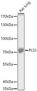
Western blot analysis of lysates from Rat lung using PLS3 Rabbit pAb (CAB3627) at 1:1000 dilution. Secondary antibody: HRP-conjugated Goat anti-Rabbit IgG (H+L) (CABS014) at 1:10000 dilution. Lysates/proteins: 25 μg per lane. Blocking buffer: 3% nonfat dry milk in TBST. Detection: ECL Basic Kit (AbGn00020). Exposure time: 30s.
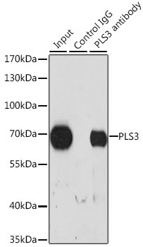
Immunoprecipitation analysis of 200 μg extracts of HeLa cells, using 3 μg PLS3 antibody (CAB3627). Western blot was performed from the immunoprecipitate using PLS3 antibody (CAB3627) at a dilution of 1:1000.