The PLD4 Antibody (CAB15207) is a high-quality antibody developed for reliable detection and analysis of target proteins. This antibody, generated in rabbits, exhibits high reactivity with human samples and has been validated for use in Western blot applications. By binding to the PLD4 protein, this antibody enables precise detection and analysis in a variety of cell types, making it ideal for studies in immunology, inflammation, and related fields.PLD4 is a key player in the regulation of inflammatory responses and immune cell activation, making it a promising target for research into autoimmune disorders, chronic inflammatory diseases, and even cancer.
This antibody is validated for use in WB, ELISA applications and has demonstrated reactivity against Human, Rat samples.
Product Name:
PLD4 Antibody
SKU:
CAB15207
Size:
20μL, 100μL
Reactivity:
Human, Rat
Conjugate:
Unconjugated
Immunogen:
Recombinant protein (or fragment).This information is considered to be commercially sensitive.
Recommended starting concentration is 1 μg/mL. Please optimize the concentration based on your specific assay requirements.
Synonyms:
C14orf175, PLD4
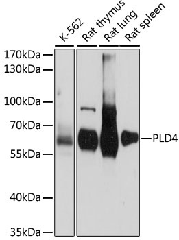
Positive Sample:
K-562, Rat thymus, Rat lung, Rat spleen
Cellular Localization:
Membrane, Single-Pass Membrane Protein.
Calculated MW:
56kDa
Observed MW:
64kDa
Predicted to enable single-stranded DNA 5'-3' exodeoxyribonuclease activity. Predicted to be involved in hematopoietic progenitor cell differentiation; phagocytosis; and regulation of cytokine production involved in inflammatory response. Predicted to be located in early endosome and endoplasmic reticulum membrane. Predicted to be active in several cellular components, including endoplasmic reticulum; phagocytic vesicle; and trans-Golgi network membrane.
Purification Method
Affinity purification
Gene ID
122618
RRID
AB_2762100
Buffer Information
Store at -20℃. Avoid freeze / thaw cycles. Buffer: PBS with 0.01% thimerosal,50% glycerol,pH7.3.
Western blot analysis of various lysates using PLD4 Rabbit pAb (CAB15207) at 1:1000 dilution. Secondary antibody: HRP-conjugated Goat anti-Rabbit IgG (H+L) (CABS014) at 1:10000 dilution. Lysates/proteins: 25μg per lane. Blocking buffer: 3% nonfat dry milk in TBST. Detection: ECL Basic Kit (AbGn00020). Exposure time: 15s.