The PLK2 Antibody (CAB7066) is a high-quality antibody developed for reliable detection and analysis of target proteins. This antibody, raised in rabbits, exhibits high reactivity with human samples and has been validated for use in Western blot applications. By binding specifically to the PLK2 protein, researchers can accurately detect and analyze its expression in a variety of cell types.PLK2, also known as polo-like kinase 2, plays a crucial role in cell division and DNA damage response, making it a key target for studies in cancer research and neurobiology.
This antibody is validated for use in WB, IF/ICC, ELISA applications and has demonstrated reactivity against Human, Mouse, Rat samples.
Product Name:
PLK2 Antibody
SKU:
CAB7066
Size:
20μL, 100μL
Reactivity:
Human, Mouse, Rat
Conjugate:
Unconjugated
Immunogen:
Recombinant protein (or fragment).This information is considered to be commercially sensitive.
The protein encoded by this gene is a member of the polo family of serine/threonine protein kinases that have a role in normal cell division. This gene is most abundantly expressed in testis, spleen and fetal tissues, and its expression is inducible by serum, suggesting that it may also play an important role in cells undergoing rapid cell division. Alternatively spliced transcript variants encoding different isoforms have been found for this gene.
Purification Method
Affinity purification
Gene ID
10769
RRID
AB_2767621
Buffer Information
Store at -20℃. Avoid freeze / thaw cycles. Buffer: PBS containing 50% glycerol, preserved with proclin300 or sodium azide, pH 7.3.
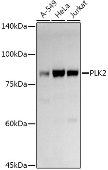
Western blot analysis of various lysates using PLK2 Rabbit pAb (CAB7066) at 1:1000 dilution. Secondary antibody: HRP-conjugated Goat anti-Rabbit IgG (H+L) (CABS014) at 1:10000 dilution. Lysates/proteins: 25μg per lane. Blocking buffer: 3% nonfat dry milk in TBST. Detection: ECL Basic Kit (AbGn00020). Exposure time: 60s.
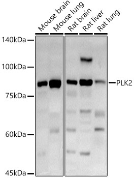
Western blot analysis of various lysates using PLK2 Rabbit pAb (CAB7066) at 1:1000 dilution. Secondary antibody: HRP-conjugated Goat anti-Rabbit IgG (H+L) (CABS014) at 1:10000 dilution. Lysates/proteins: 25μg per lane. Blocking buffer: 3% nonfat dry milk in TBST. Detection: ECL Basic Kit (AbGn00020). Exposure time: 180s.
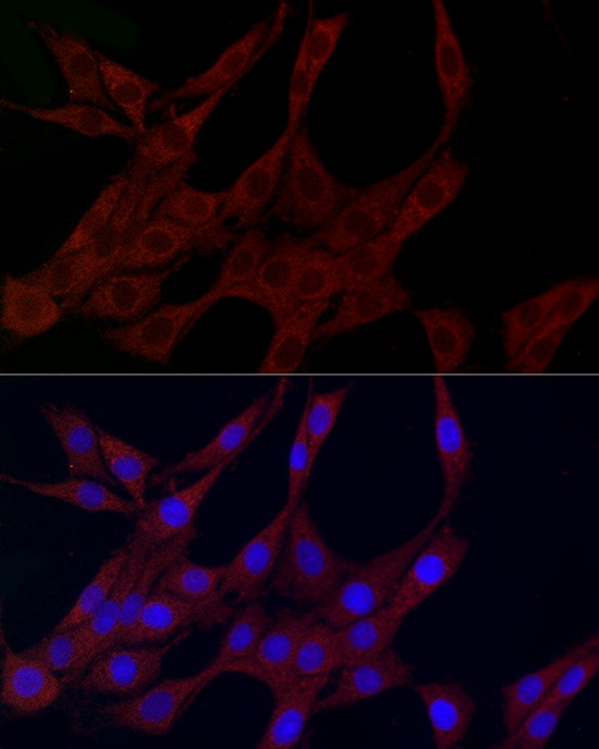
Immunofluorescence analysis of PC-12 cells using PLK2 Rabbit pAb (CAB7066) at dilution of 1:100 (40x lens). Secondary antibody: Cy3-conjugated Goat anti-Rabbit IgG (H+L) (CABS007) at 1:500 dilution. Blue: DAPI for nuclear staining.