The PLK3 Antibody (CAB8674) is a high-quality antibody developed for reliable detection and analysis of target proteins. This antibody, produced in rabbits, exhibits high reactivity with human samples and is validated for use in various applications including Western blotting.PLK3, also known as Polo-like kinase 3, plays a critical role in DNA damage-induced cell cycle arrest and apoptosis. Its involvement in these processes makes it a potential target for cancer therapy and research into genetic disorders. By targeting PLK3, researchers can better understand its role in cell cycle progression and potentially develop novel therapeutic strategies for cancer treatment.
This antibody is validated for use in WB, ELISA applications and has demonstrated reactivity against Human, Mouse, Rat samples.
Product Name:
PLK3 Antibody
SKU:
CAB8674
Size:
20μL, 100μL
Reactivity:
Human, Mouse, Rat
Conjugate:
Unconjugated
Immunogen:
Recombinant protein (or fragment).This information is considered to be commercially sensitive.
Recommended starting concentration is 1 μg/mL. Please optimize the concentration based on your specific assay requirements.
Synonyms:
CNK, FNK, PRK, PLK-3, PLK3
Positive Sample:
HeLa, Mouse large intestine, Rat brain
Cellular Localization:
Cytoplasm, Golgi Apparatus, Nucleus, Centrosome, Cytoskeleton, Microtubule Organizing Center, Nucleolus.
Calculated MW:
72kDa
Observed MW:
72kDa
The protein encoded by this gene is a member of the highly conserved polo-like kinase family of serine/threonine kinases. Members of this family are characterized by an amino-terminal kinase domain and a carboxy-terminal bipartite polo box domain that functions as a substrate-binding motif and a cellular localization signal. Polo-like kinases are important regulators of cell cycle progression. This gene has also been implicated in stress responses and double-strand break repair. In human cell lines, this protein is reported to associate with centrosomes in a microtubule-dependent manner, and during mitosis, the protein becomes localized to the mitotic apparatus. Expression of a kinase-defective mutant results in abnormal cell morphology caused by changes in microtubule dynamics and mitotic arrest followed by apoptosis.
Purification Method
Affinity purification
Gene ID
1263
RRID
AB_2771713
Buffer Information
Store at -20℃. Avoid freeze / thaw cycles. Buffer: PBS containing 50% glycerol, preserved with proclin300 or sodium azide, pH 7.3.
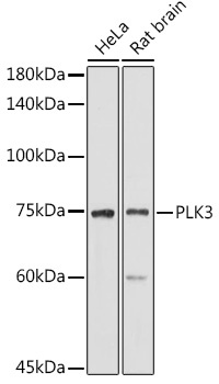
Western blot analysis of various lysates using PLK3 Rabbit pAb (CAB8674) at 1:1000 dilution. Secondary antibody: HRP-conjugated Goat anti-Rabbit IgG (H+L) (CABS014) at 1:10000 dilution. Lysates/proteins: 25μg per lane. Blocking buffer: 3% nonfat dry milk in TBST. Detection: ECL Basic Kit (AbGn00020). Exposure time: 180s.
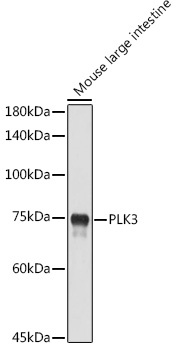
Western blot analysis of lysates from Mouse large intestine, using PLK3 Rabbit pAb (CAB8674) at 1:1000 dilution. Secondary antibody: HRP-conjugated Goat anti-Rabbit IgG (H+L) (CABS014) at 1:10000 dilution. Lysates/proteins: 25μg per lane. Blocking buffer: 3% nonfat dry milk in TBST. Detection: ECL Enhanced Kit (AbGn00021). Exposure time: 180s.