The PLP1 Antibody (CAB20009) is a high-quality antibody developed for reliable detection and analysis of target proteins. This gene encodes a transmembrane proteolipid protein that is the predominant component of myelin. The encoded protein may play a role in the compaction, stabilization, and maintenance of myelin sheaths, as well as in oligodendrocyte development and axonal survival. Mutations in this gene cause Pelizaeus-Merzbacher disease and spastic paraplegia type 2. Alternatively splicing results in multiple transcript variants, including the DM20 splice variant.
This antibody is validated for use in WB, IHC-P, ELISA, IF-P applications and has demonstrated reactivity against Human, Mouse, Rat samples.
Product Name:
PLP1 Antibody
SKU:
CAB20009
Size:
100μL, 20μL
Reactivity:
Human, Mouse, Rat
Conjugate:
Unconjugated
Immunogen:
Recombinant protein (or fragment).This information is considered to be commercially sensitive.
Tested Applications:
WBIHC-PELISAIF-P
Recommended Dilution:
WB
1:1000 - 1:5000
IF-P
1:50 - 1:200
IHC-P
1:50 - 1:200
ELISA
Recommended starting concentration is 1 μg/mL. Please optimize the concentration based on your specific assay requirements.
This gene encodes a transmembrane proteolipid protein that is the predominant component of myelin. The encoded protein may play a role in the compaction, stabilization, and maintenance of myelin sheaths, as well as in oligodendrocyte development and axonal survival. Mutations in this gene cause Pelizaeus-Merzbacher disease and spastic paraplegia type 2. Alternatively splicing results in multiple transcript variants, including the DM20 splice variant.
Purification Method
Affinity purification
Gene ID
5354
RRID
AB_2862916
Buffer Information
Store at -20℃. Avoid freeze / thaw cycles. Buffer: PBS containing 50% glycerol, preserved with proclin300 or sodium azide, pH 7.3.
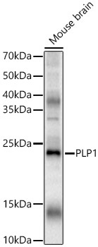
Western blot analysis of various lysates, using PLP1 Rabbit pAb (CAB20009) at 1:2000 dilution. Secondary antibody: HRP-conjugated Goat anti-Rabbit IgG (H+L) (AS014) at 1:10000 dilution. Lysates/proteins: 25μg per lane. Blocking buffer: 3% nonfat dry milk in TBST. Detection: ECL Basic Kit (AbGn00020). Exposure time: 30s.
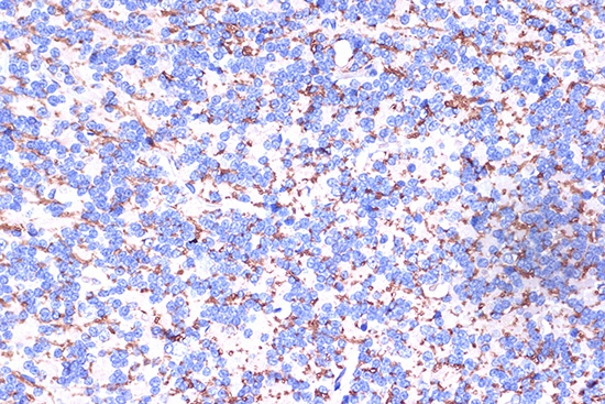
Immunohistochemistry analysis of paraffin-embedded Rat brain using PLP1 Rabbit pAb (CAB20009) at dilution of 1:100 (40x lens). Microwave antigen retrieval performed with 0.01M PBS Buffer (pH 7.2) prior to IHC staining.
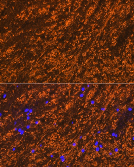
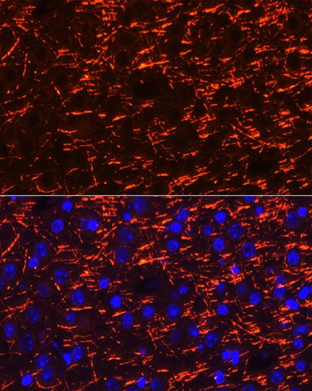
Immunofluorescence analysis of paraffin-embedded rat brain using PLP1 Rabbit pAb (CAB20009) at dilution of 1:100 (40x lens). Secondary antibody: Cy3-conjugated Goat anti-Rabbit IgG (H+L) (AS007) at 1:500 dilution. Blue: DAPI for nuclear staining.
Immunofluorescence analysis of paraffin-embedded mouse brain using PLP1 Rabbit pAb (CAB20009) at dilution of 1:100 (40x lens). Secondary antibody: Cy3-conjugated Goat anti-Rabbit IgG (H+L) (AS007) at 1:500 dilution. Blue: DAPI for nuclear staining.