The PLS1 Antibody (CAB15303) is a high-quality antibody developed for reliable detection and analysis of target proteins. This antibody, generated in rabbits, exhibits high reactivity with human samples and has been validated for use in Western blot applications. By binding specifically to the PLS1 protein, this antibody enables accurate detection and analysis in various cell types, making it ideal for studies in cell biology and cancer research.PLS1, also known as plastin 1, is a cytoskeletal protein that plays a crucial role in regulating the movement and shape of cells.
This antibody is validated for use in WB, IHC-P, ELISA applications and has demonstrated reactivity against Human, Mouse, Rat samples.
Product Name:
PLS1 Antibody
SKU:
CAB15303
Size:
20μL, 100μL
Reactivity:
Human, Mouse, Rat
Conjugate:
Unconjugated
Immunogen:
Recombinant protein (or fragment).This information is considered to be commercially sensitive.
Recommended starting concentration is 1 μg/mL. Please optimize the concentration based on your specific assay requirements.
Synonyms:
DFNA76, PLS1
Positive Sample:
HT-29, 293T, LO2, Mouse brain, Mouse liver, Rat spleen
Cellular Localization:
Cytoplasm.
Calculated MW:
70kDa
Observed MW:
70kDa
Plastins are a family of actin-binding proteins that are conserved throughout eukaryote evolution and expressed in most tissues of higher eukaryotes. In humans, two ubiquitous plastin isoforms (L and T) have been identified. The protein encoded by this gene is a third distinct plastin isoform, which is specifically expressed at high levels in the small intestine. Alternatively spliced transcript variants varying in the 5' UTR, but encoding the same protein, have been found for this gene. A pseudogene of this gene is found on chromosome 11.
Purification Method
Affinity purification
Gene ID
5357
RRID
AB_2762205
Buffer Information
Store at -20℃. Avoid freeze / thaw cycles. Buffer: PBS with 0.01% thimerosal,50% glycerol,pH7.3.
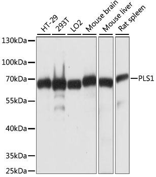
Western blot analysis of various lysates using PLS1 Rabbit pAb (CAB15303) at 1:1000 dilution. Secondary antibody: HRP-conjugated Goat anti-Rabbit IgG (H+L) (CABS014) at 1:10000 dilution. Lysates/proteins: 25μg per lane. Blocking buffer: 3% nonfat dry milk in TBST. Detection: ECL Basic Kit (AbGn00020). Exposure time: 3s.
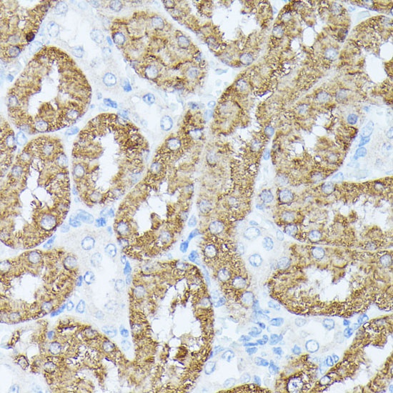
Immunohistochemistry analysis of paraffin-embedded Rat kidney using PLS1 Rabbit pAb (CAB15303) at dilution of 1:100 (40x lens). Microwave antigen retrieval performed with 0.01M PBS Buffer (pH 7.2) prior to IHC staining.